The bones of the foot (ossa pedis) include the tarsal bones (ossa tarsi), the metatarsals (ossa metatarsi), and the phalanges (phalanges/ossa digitorum).
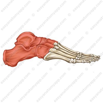
Let us examine the tarsal bones. These are 7 small bones that form two rows — a proximal and a distal row.
The bones of the proximal row are the following:
talus (talus)

calcaneus (calcaneus)
Calcaneus (calcaneus) 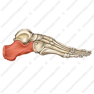
Now come the bones of the distal row:
navicular (os naviculare)
Navicular (os naviculare) 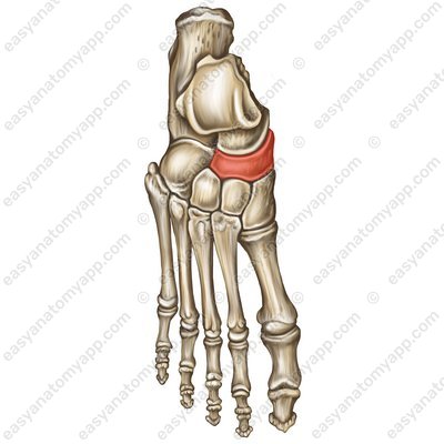
cuboid (os cuboideum)
Cuboid (os cuboideum) 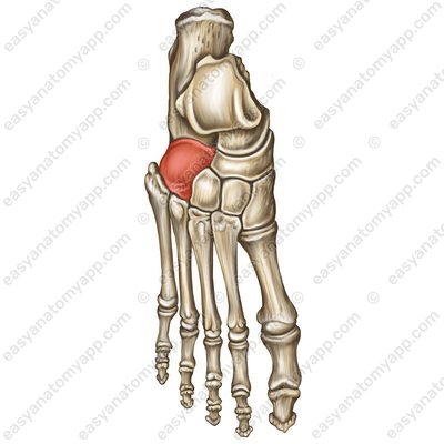
medial cuneiform (os cuneiforme mediale)
Medial cuneiform (os cuneiforme mediale) 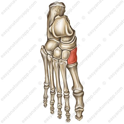
intermediate cuneiform (os cuneiforme intermedium)
Intermediate cuneiform (os cuneiforme intermedium) 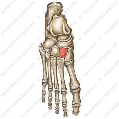
lateral cuneiform (os cuneiforme laterale)
Lateral cuneiform (os cuneiforme laterale) 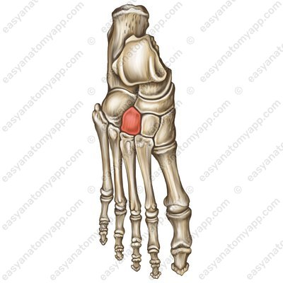
The talus consists of several parts:
body (corpus tali)
Body (corpus tali) head (caput tali)
Head (caput tali) 
neck (collum tali)
Neck (collum tali) 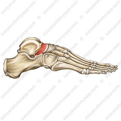
On the body of the bone is the trochlea of the talus (trochlea tali).
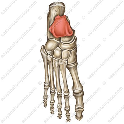
This trochlea has three articular surfaces:
superior surface (facies superior), connecting to the inferior articular surface of the tibia
Superior surface (facies superior) 
medial malleolar facet (facies malleolaris medialis), connecting to the ankle surface of the tibia
Medial malleolar facet (facies malleolaris medialis) 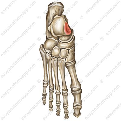
lateral malleolar facet (facies malleolaris lateralis), connecting to the ankle surface of the fibula
Lateral malleolar facet (facies malleolaris lateralis), 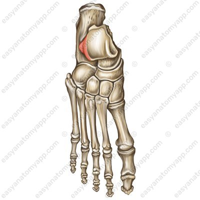
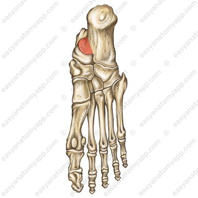
The posterior process of the talus (processus posterior tali) is derived from the body of the bone.
Posterior process of the talus (processus posterior tali) 
This process has a groove for the tendon for the flexor hallucis longus (sulcus tendinis musculi flexoris hallucis longi), which divides the process into a medial tubercle (tuberculum mediale)
Tendon for the flexor hallucis longus (sulcus tendinis musculi flexoris hallucis longi) 
Medial tubercle (tuberculum mediale) 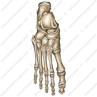
and a lateral tubercle (tuberculum laterale).
Lateral tubercle (tuberculum laterale) 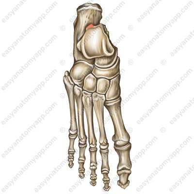
The lower part of the talus also has three articular surfaces that connect to the calcaneus:
the anterior facet for the calcaneus (facies articularis calcanea anterior)
the middle facet for the calcaneus (facies articularis calcanea media)
the posterior facet for the calcaneus (facies articularis calcanea posterior)
Between the posterior and middle facets lies talar sulcus (sulcus tali), to which the ligaments are attached.
The head of the talus has a navicular articular surface (facies articularis navicularis) that connects to the navicular.
The calcaneus is under the talus.
Calcaneus At the back of the calcaneus lies the calcaneal tuberosity (tuber calcanei).
Calcaneal tuberosity (tuber calcanei) 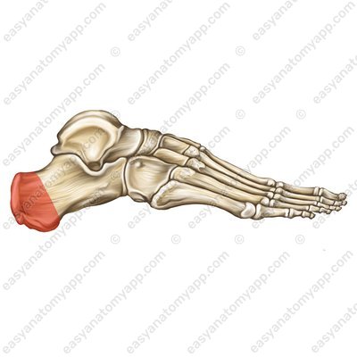
From the calcaneal tuberosity the lateral process of the calcaneal tuberosity (processus lateralis tuberis calcanei)
Lateral process of the calcaneal tuberosity (processus lateralis tuberis calcanei) 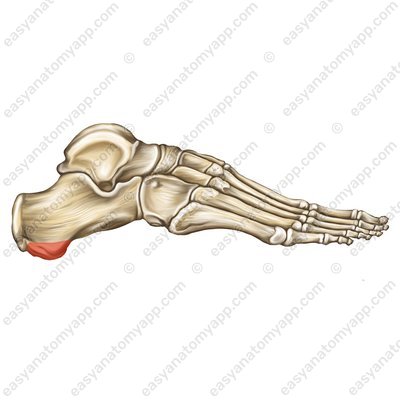
and the medial process of the calcaneal tuberosity (processus medialis tuberis calcanei) depart.
Medial process of the calcaneal tuberosity (processus medialis tuberis calcanei) 
The upper part of the calcaneus also has three articular surfaces that connect to the talus:
the anterior talar articular surface (facies articularis talaris anterior)
the medial talar articular surface (facies articularis talaris media)
the posterior talar articular surface (facies articularis talaris posterior)
Between the posterior and middle talar articular surfaces lies a calcaneal sulcus (sulcus calcanei). Together with the corresponding talar sulcus, it forms the tarsal sinus (sinus tarsi), in which there is a ligament that connects these two bones.

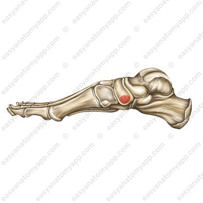
On the medial side, the so-called sustentaculum tali is derived from the talus.
On the front part of the bone there is an articular surface for the cuboid (facies articularis cuboidea), which connects to the cuboid.
On the lateral surface there is the groove for the tendon of the fibularis longus (sulcus tendinis musculi fibularis longi).
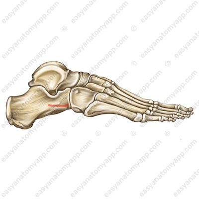
On its lateral surface there is also the fibular trochlea (trochlea fibularis).
.jpg)
On the navicular, the tuberosity of the navicular (tuberositas ossis navicularis) can be distinguished.
And on the cuboid we can see the tuberosity of the cuboid (tuberositas ossis cuboidei).

There are no specific formations on the cuneiform bones, which should be separately mentioned.
Tarsal bones
- bones of foot
- ossa pedis
- tarsal bones
- ossa tarsi
- metatarsals
- ossa metatarsi
- phalanges
- phalanges/ossa digitorum
- talus
- talus
- calcaneus
- calcaneus
- navicular
- os naviculare
- cuboid
- os cuboideum
- medial cuneiform
- os cuneiforme mediale
- intermediate cuneiform
- os cuneiforme intermedium
- lateral cuneiform
- os cuneiforme laterale
- body of talus
- corpus tali
- head of talus
- caput tali
- neck of talus
- collum tali
- trochlea of talus
- trochlea tali
- superior surface
- facies superior
- medial malleolar facet
- facies malleolaris medialis
- lateral malleolar facet
- facies malleolaris lateralis
- posterior process of talus
- processus posterior tali
- groove for tendon for flexor hallucis longus
- sulcus tendinis musculi flexoris hallucis longi
- medial tubercle
- tuberculum mediale
- lateral tubercle
- tuberculum laterale
- anterior facet for calcaneus
- facies articularis calcanea anterior
- middle facet for calcaneus
- facies articularis calcanea media
- posterior facet for calcaneus
- facies articularis calcanea posterior
- talar sulcus
- sulcus tali
- navicular articular surface
- facies articularis navicularis
- calcaneal tuberosity
- tuber calcanei
- lateral process of calcaneal tuberosity
- processus lateralis tuberis calcanei
- medial process of calcaneal tuberosity
- processus medialis tuberis calcanei
- anterior talar articular surface
- facies articularis talaris anterior
- middle talar articular surface
- facies articularis talaris media
- posterior talar articular surface
- facies articularis talaris posterior
- calcaneal sulcus
- sulcus calcanei
- tarsal sinus
- sinus tarsi
- sustentaculum tali
- sustentaculum tali
- articular surface for cuboid
- facies articularis cuboidea
- groove for tendon of fibularis longus
- sulcus tendinis musculi fibularis longi
- fibular trochlea
- trochlea fibularis
- tuberosity of navicular
- tuberositas ossis navicularis
- tuberosity of cuboid
- tuberositas ossis cuboidei

