Let us examine the structure of the five sacral vertebrae (vertebrae sacrales). They are fused into a single triangular bone — the sacrum (os sacrum).
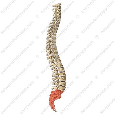
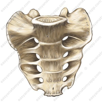
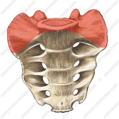
The following parts are distinguished in the sacrum:
Base of the sacrum (basis ossis sacri)
Lateral part (pars lateralis)
Apex of the sacrum (apex ossis sacri)
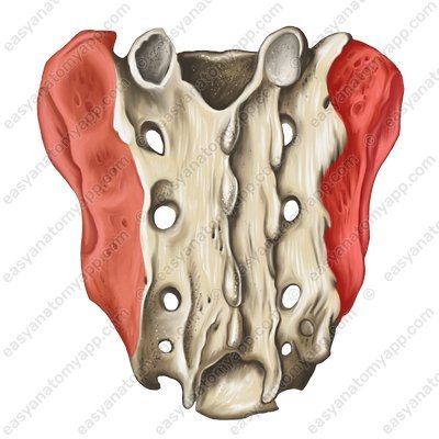
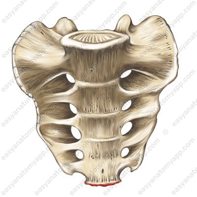
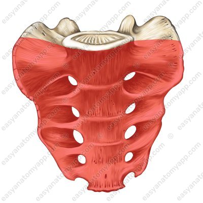
There are also two surfaces — the pelvic surface (facies pelvica) and the dorsal surface (facies dorsalis).
.jpg)
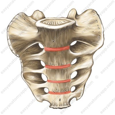
On the pelvic surface, at the junction of the sacrum with the fifth lumbar vertebrae, the so-called promontory (promontorium) comes forward.
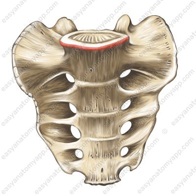
Below you can see the transverse ridges (lineae transversae) — traces of the fusion of the bodies of the sacral vertebrae.
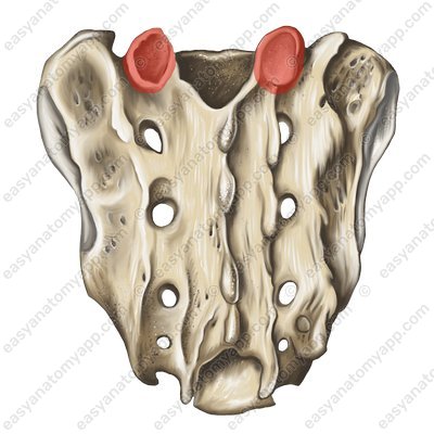
And on the dorsal surface on each side is the superior articular process (processus articularis superior), with a corresponding superior articular surface (facies articularis superior) for articulation with the fifth lumbar vertebra.
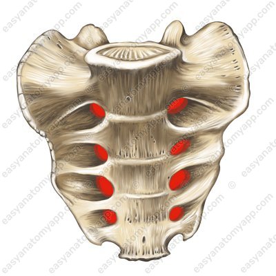
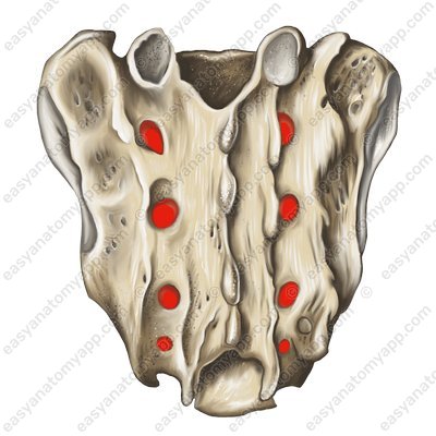
On both surfaces of the sacrum are the anterior sacral foramina (foramina sacralia anteriora), through which the anterior branches of the sacral nerves and blood vessels pass, and the posterior sacral foramina (foramina sacralia posteriora), through which the posterior branches of the sacral nerves and blood vessels pass.
Moreover, the vertebral foramen (foramen vertebrale) of the sacral vertebrae form the sacral canal (canalis sacralis).
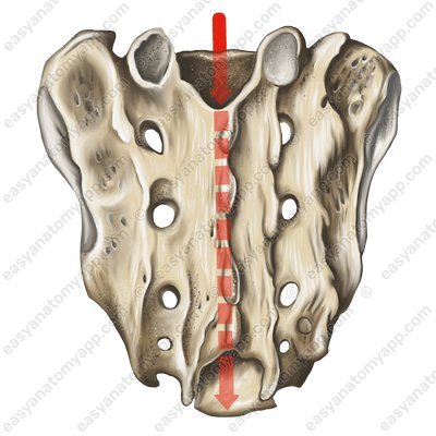
At the apex, the entrance to this canal is called the sacral hiatus (hiatus sacralis).
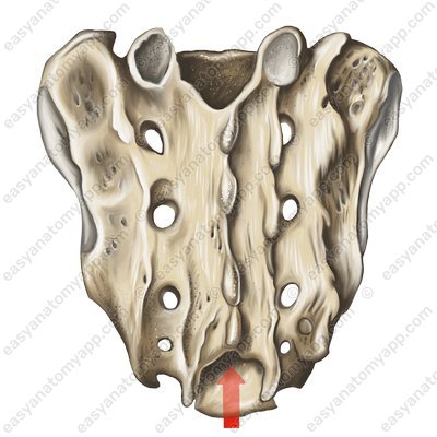
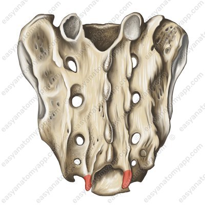
This hiatus is bounded on each side by the sacral horn (cornu sacrale).
On the dorsal surface of the sacrum, there are longitudinal crests, which are the result of the fusion of the vertebral processes:
The median sacral crest (crista sacralis mediana) is a trace of the fusion of the spinous processes
The median sacral crest (crista sacralis mediana) 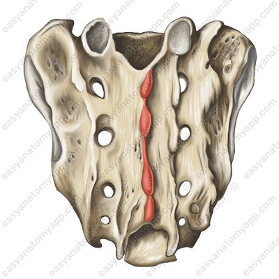
The intermediate sacral crest (crista sacralis medialis) is a trace of the fusion of the articular processes
The intermediate sacral crest (crista sacralis medialis) 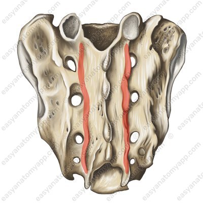
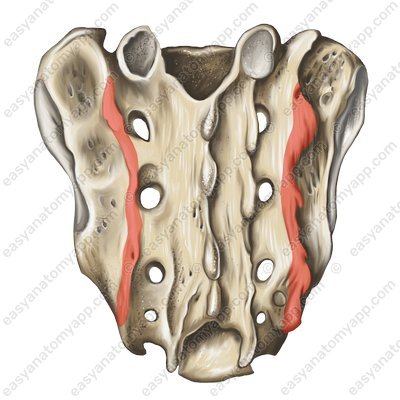
The lateral sacral crest (crista sacralis lateralis) is a trace of fusion of the transverse processes
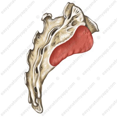
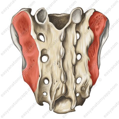
On the lateral parts of the sacrum, there are auricular surfaces (facies auricularis), which articulate with the pelvic bones
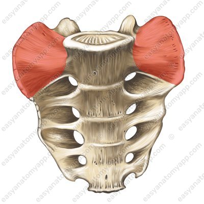
The anterior surface of the lateral part is called the ala of the sacrum (ala ossis sacri), and on the posterior is the sacral tuberosity (tuberositas ossis sacri), where the ligaments are attached.
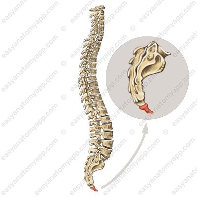
We now come to the structure of the coccygeal vertebrae (vertebrae coccygeae). Their number is not constant (1-5, most often 3).
Like the sacrum, they are fused into a single bone — the coccyx (os coccygis). The coccyx has a base (basis), an apex (apex), as well as a coccygeal cornu (cornu coccygeum), which is used on each side to articulate with the sacrum.
Sacrum & Coccyx
- sacral vertebrae
- vertebrae sacrales
- sacrum
- os sacrum
- base of sacrum
- basis ossis sacri
- lateral part
- pars lateralis
- apex of sacrum
- apex ossis sacri
- pelvis surface
- facies pelvica
- dorsal surface
- facies dorsalis
- promontory
- promontorium
- transverse ridges
- lineae transversae
- superior articular process
- processus articularis superior
- superior articular facet
- facies articularis superior
- anterior sacral foramina
- foramina sacralia anteriora
- posterior sacral foramina
- foramina sacralia posteriora
- vertebral foramen
- foramen vertebrale
- sacral canal
- canalis sacralis
- sacral hiatus
- hiatus sacralis
- sacral horn
- cornu sacrale
- median sacral crest
- crista sacralis mediana
- intermediate sacral crest
- crista sacralis medialis
- lateral sacral crest
- crista sacralis lateralis
- auricular surface
- facies auricularis
- ala of sacrum
- ala ossis sacri
- sacral tuberosity
- tuberositas ossis sacri
- coccygeal vertebrae
- vertebrae coccygeae
- coccyx
- os coccygis
- coccygeal cornu
- cornu coccygeum

