In addition to the ankle joint, which is discussed in a separate video, the joints of the foot include several more groups: the joints of the tarsal bones, the joints between the tarsal and metatarsal bones, and the joints of the phalanges.
Let’s learn about the last two groups.
Let’s start with the joints that connect the tarsal bones with the metatarsal bones.
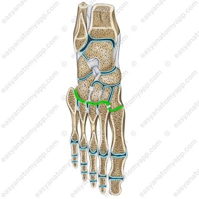
The tarsometatarsal joints (articulationes tarsometatarseae). The combination of these joints is called the Lisfranc joint.
.jpg)
.jpg)
The bones forming the joint are medial cuneiform bone (os cuneiforme mediale), intermediate cuneiform bone(os cuneiforme intermedium), lateral cuneiform bone (os cuneiforme laterale), cuboid bone(os cuboideum) and the 1st-5th metatarsals (ossa metatarsalia I-V).
The articular surfaces involved in the formation of the joint are the distal articular surfaces of the cuneiform bones, the articular surface of the cuboid bone and the articular surfaces of the bases of the metatarsal bones.
In this case, three isolated joints are formed:
The joint between the medial cuneiform bone and the 1st metatarsal bone
The joint between the 2nd and the 3rd metatarsals and the intermediate and lateral cuneiform bones
The joint between the cuboid bone and the 4th and 5th metatarsals
The joint capsule is attached along the edges of the articular surfaces.
According to the classification, this joint is plane.
Almost no motions take place in this joint.
The ligamentous apparatus of the joint consists of several ligaments.
The dorsal tarsometatarsal ligaments (ligamenta tarsometatarsea dorsalia)
Dorsal tarsometatarsal ligaments (ligg. tarsometatarsalia dorsalia) 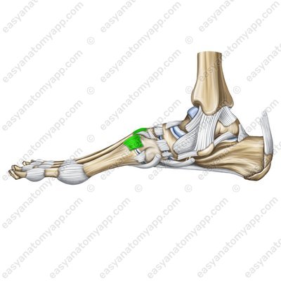
The plantar tarsometatarsal ligaments (ligamenta tarsometatarsea plantaria)
Plantar tarsometatarsal ligaments (ligg. tarsometatarsalia plantaria) 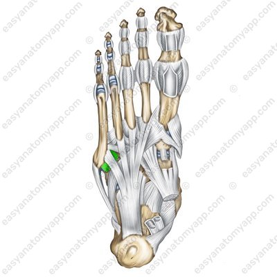
Plantar tarsometatarsal ligaments (ligg. tarsometatarsalia plantaria) 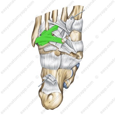
The interosseous cuneometatarsal ligaments (ligamenta cuneometatarsea interossea). The medial cuneometatarsal ligament, which connects the medial cuneometatarsal bone with the 2nd metatarsal is called the key of the Lisfranc joint.
Interosseous cuneometatarsal ligaments (ligg. cuneometatarseae interossea) 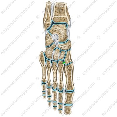
The intermetatarsal joints (articulationes intermetatarseae).
The bones forming the joint are the 1st-5th metatarsals (ossa metatarsalia I-V).
Intermetatarsal joints (artt. intermetatarsales) .jpg)
The articular surfaces involved in the formation of the joint are the surfaces of the metatarsals facing each other.
The joint capsules are attached along the edges of the articular surfaces.
The metatarsal interosseous ligaments (ligamenta metatarsea interossea) belong to the ligamentous apparatus of the joint.
Metatarsal interosseous ligaments (ligamenta metatarsea interossea) 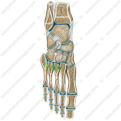
Dorsal metatarsal ligaments (ligg. metatarsalia dorsalia)
Dorsal metatarsal ligaments (ligg. metatarsalia dorsalia) .jpg)
Plantar metatarsal ligaments (ligg. metatarsalia plantaria)
Plantar metatarsal ligaments (ligg. metatarsalia plantaria) 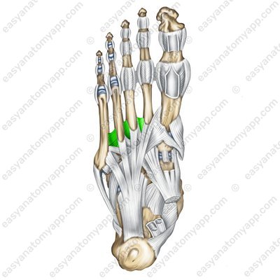
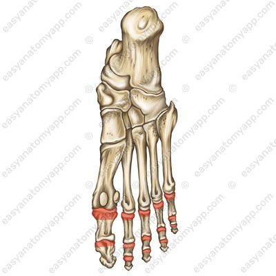
The metatarsophalangeal joints (articulationes metatarsophalangeae)
Metatarsophalangeal joints (artt. metatarsophalangeae) .jpg)
The bones forming the joint are the 1st-5th proximal phalanges of the toes (phalanx proximalis) and the 1st-5th metatarsals (ossa metatarsalia I-V).
The articular surfaces involved in the formation of the joint are the articular surfaces of the heads of the metatarsal bones and the articular surfaces of the bases of the proximal phalanges.
Head of the metatarsal bones (caput ossis metatarsi) 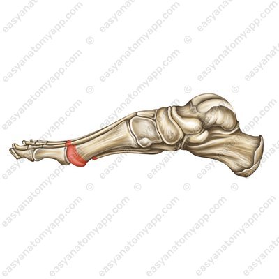
Bases of the proximal phalanges (basis phalangis) 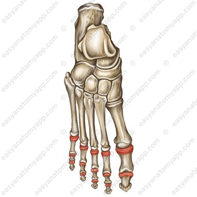
The joint capsule is very thin and loose, it is attached along the edges of the articular surfaces.
According to the classification, the joints are ellipsoid and biaxial.
Flexion and extension of the toes, as well as their slight adduction and abduction are carried out in the joint.
The fixing apparatus of the joint is composed of several ligaments:
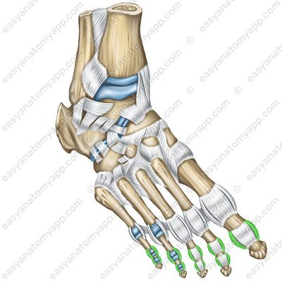
These are the lateral and medial collateral ligaments (ligamenta collateralia mediales et laterales)
Collateral ligaments (ligg. collateralia) 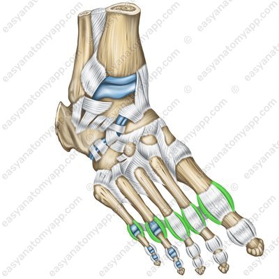
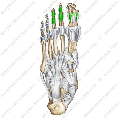
The plantar ligaments (ligamenta plantaria)
And the deep transverse metatarsal ligament (ligamentum metatarseum transversum profundum) passes transversely from the heads of the 1st to the head of the 5th of the metatarsals, fusing with the capsules of the metatarsal joints and connecting the heads of all metatarsal bones.
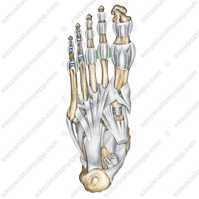
The interphalangeal joints of the foot (articulationes interphalangeae pedis)
The bones forming the joint are the phalanges of the toes (phalanges)
The articular surfaces involved in the formation of the joint are the articular surface on the head of one phalanx (caput phalangis) and the articular surface on the base of the other phalanx (basis phalangis).
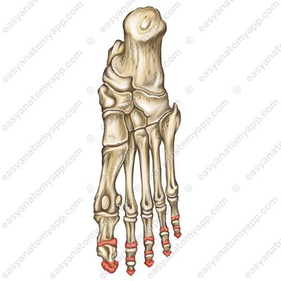
The joint capsules are attached along the edges of the articular surfaces.
According to the classification, the joints are hinge and uniaxial.
Flexion and extension are carried out in the interphalangeal joints. At the same time, the motions of the thumb have a large amplitude.
The ligamentous apparatus of the joint includes collateral ligaments (ligamenta collateralia) and plantar ligaments (ligamenta plantari).
Joint of the foot
- Tarsometatarsal joints
- articulationes tarsometatarseae
- Medial cuneiform bone
- os cuneiforme mediale
- Intermediate cuneiform bone
- os cuneiforme intermedium
- Lateral cuneiform bone
- os cuneiforme laterale
- Cuboid bone
- os cuboideum
- The 1st-5th metatarsals
- ossa metatarsalia I-V
- Dorsal tarsometatarsal ligaments
- ligamenta tarsometatarsea dorsalia
- Plantar tarsometatarsal ligaments
- ligamenta tarsometatarsea plantaria
- Interosseous cuneometatarsal ligaments
- ligamenta cuneometatarsea interossea
- Intermetatarsal joints
- articulationes intermetatarseae
- Metatarsal interosseous ligaments
- ligamenta metatarsea interossea
- Metatarsophalangeal joints
- articulationes metatarsophalangeae
- Proximal phalanges
- phalanx proximalis
- Lateral and medial collateral ligaments
- ligamenta collateralia mediales et laterales
- Plantar ligaments
- ligamenta plantaria
- Deep transverse metatarsal ligament
- ligamentum metatarseum transversum profundum
- Interphalangeal joints of foot
- articulationes interphalangeae pedis
- Phalanges of the toes
- phalanges
- Head of the phalanx
- caput phalangis
- Base of the phalanx
- basis phalangis
- Collateral ligaments
- ligamenta collateralia
- Plantar ligaments
- ligamenta plantari

