The vertebral column is connected to the skull by three joints: the atlanto-occipital, the median atlanto-axial, and the lateral atlanto-axial joint.
Let’s learn about the structure of the lateral atlanto-axial joint (articulatio atlantoaxialis lateralis).
.jpg)
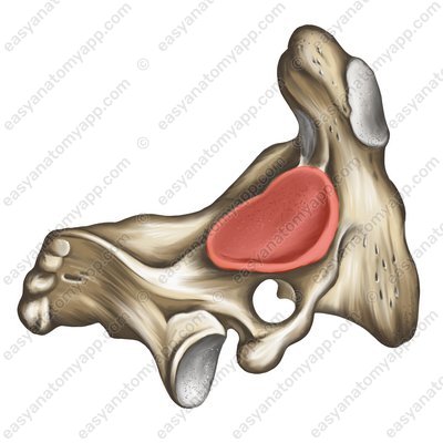
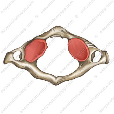
This joint is formed between the inferior articular surface of the atlas (facies articularis inferior) and the articular surface on the superior articular process of the axis (facies articularis superior).
The joint capsule is attached along the edge of the articular surfaces.
According to the classification, this joint is plane, multiaxial, simple, and combined (with the same joint of the opposite side, as well as with the median atlanto-axial joint).
The lateral atlanto-axial joint is restricted in mobility, sliding motions are carried out in it only with a slight displacement of the articular surfaces relative to each other.
As a rule, this takes place around the vertical axis, while the head turns to the right and left.
The ligamentous apparatus of the joint includes several ligaments:
The cruciate ligament of the atlas (ligamentum cruciforme atlantis). It consists of three bundles:
Cruciate ligament of the atlas (lig. cruciforme atlantis) 
The superior longitudinal fasciculus (fasciculus longitudinalis superior)
Superior longitudinal fasciculus (fasciculus longitudinalis superior) 
The inferior longitudinal fasciculus (fasciculus longitudinalis inferior)
Inferior longitudinal fasciculus (fasciculus longitudinalis inferior) 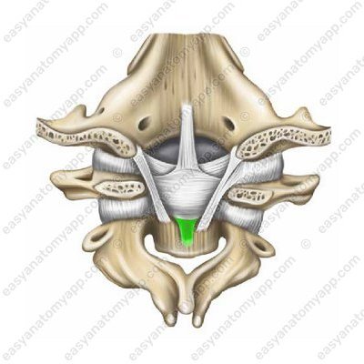
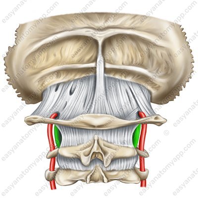
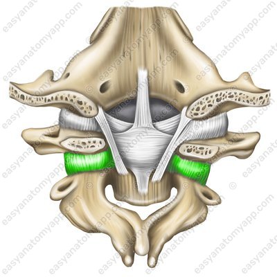
The transverse ligament of the atlas (ligamentum transversum atlantis)
Transverse ligament of the atlas (lig. transversum atlantis) 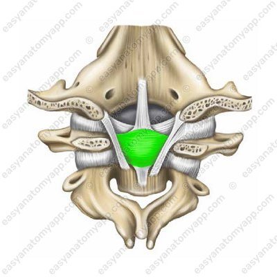
The joint is also strengthened by the alar ligaments (ligamenta alaria), which restrict excessive rotation of the head to the right and left. Each such ligament arises from the lateral surface of the dens and passes externally, obliquely and upward, where it inserts into the internal surface of the corresponding condyle of the occipital bone.
Alar ligaments (ligg. alaria) 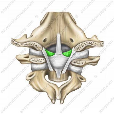
Another ligament is the apical ligament of the dens (ligamentum apicis dentis), it is unpaired, thin, and stretched between the edge of the foramen magnum and the apex of the dens.
Apical ligament of the dens (lig. apicis dentis) 
And the last ligament that strengthens the joint is the tectorial membrane (membrana tectoria).
Tectorial membrane (membrana tectoria) 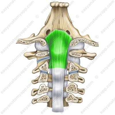
The following vessels and nerves take part in the blood supply and innervation of the joint.
Arteries: branches of the deep cervical, occipital, and vertebral arteries
Nerves: anterior branch of the second spinal nerve
Lateral atlanto-axial joint
- lateral atlanto-axial joint
- articulatio atlantoaxialis lateralis
- inferior articular surface
- facies articularis inferior
- superior articular surface
- facies articularis superior
- cruciate ligament of atlas
- ligamentum cruciforme atlantis
- superior longitudinal fasciculus
- fasciculus longitudinalis superior
- inferior longitudinal fasciculus
- fasciculus longitudinalis inferior
- transverse ligament of atlas
- ligamentum transversum atlantis
- alar ligaments
- ligamenta alaria
- apical ligament of dens
- ligamentum apicis dentis
- tectorial membrane
- membrana tectoria

