In this pdf-note, we’ll start with the borders of the lower limb:
The posterior border passes through the coccyx and the lateral margins of the sacrum
The lateral border is the iliac crest
The anterior border passes through the inguinal ligament and the pubic symphysis
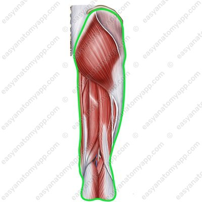
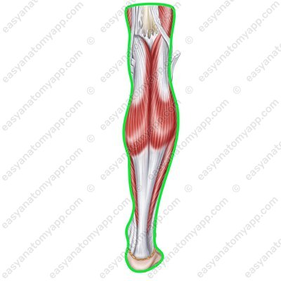
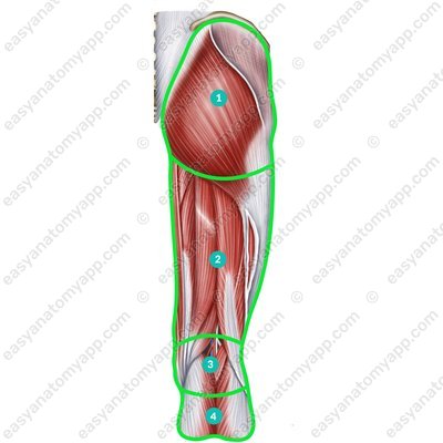
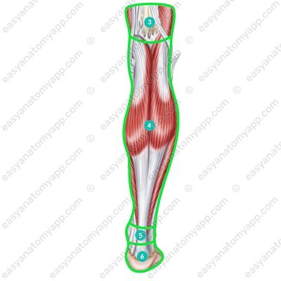
There are several regions within the lower limb:
Gluteal region (regio glutealis)
Femoral region (regio femoralis)
Knee region (regio genus)
Leg region (regio cruralis)
Ankle region (regio talocruralis)
Foot region (regio pedis)
Within these areas, the muscles of the lower limb are located.
These include:
Pelvic girdle muscles
Muscles of the free part of lower limb
The muscles of the free part of the lower limb are divided into:
Thigh muscles
Leg muscles
Foot muscles
The leg muscles form three groups:
The anterior group consists of the extensors of the foot
The posterior group consists of the flexors and supinators of the foot
The lateral group consists of the foot abductor muscles The anterior group includes the following muscles:
Tibialis anterior muscle (m. tibialis anterior)
Tibialis anterior muscle (m. tibialis anterior) 
Tibialis anterior muscle (m. tibialis anterior) 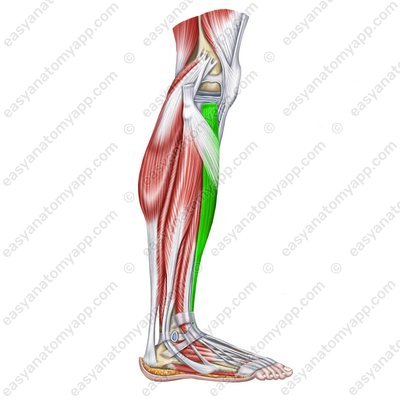

Tibialis anterior muscle (m. tibialis anterior) Tibialis anterior muscle
(m. tibialis anterior)Origin: lateral surface of the body of the tibia, lateral condyle of the tibia, interosseous membrane of the leg
Insertion: plantar surface of the medial cuneiform bone, base of the 1st metatarsal
Function: extends the foot at the ankle joint (the so-called dorsal flexion), supinates the foot
Innervation: deep fibular nerve (L4-S1)
Blood supply: anterior tibial artery
Extensor digitorum longus muscle (m. extensor digitorum longus)
Extensor digitorum longus muscle (m. extensor digitorum longus) 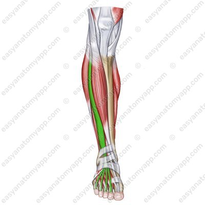
Extensor digitorum longus muscle (m. extensor digitorum longus) 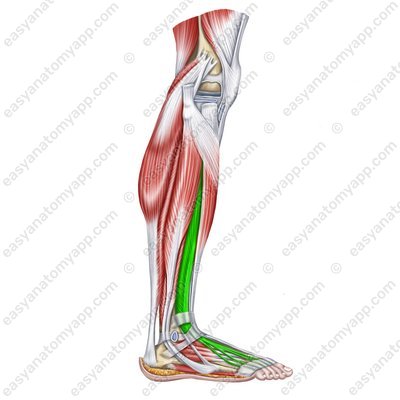
Extensor digitorum longus muscle (m. extensor digitorum longus) Extensor digitorum longus muscle
(m. extensor digitorum longus)Origin: lateral condyle of the tibia, head and anterior margin of the fibula, interosseous membrane of the leg
Insertion: at the level of the ankle joint, it divides into four tendons, each of which then divide into three bundles. The middle bundle inserts into the base of the middle phalanx, and the lateral ones insert into the base of the distal phalanx. Sometimes the muscle also has a fifth tendon.
Function: extends fingers 2-5 at the metatarsophalangeal joints, extends the foot at the ankle joint
Innervation: deep fibular nerve (L4-S1)
Blood supply: anterior tibial artery
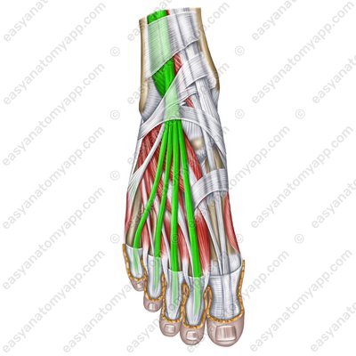
Peroneus tertius muscle (m. peroneus tertius), which is not always present
Peroneus tertius muscle (m. peroneus tertius) – tendon  2.jpg)
.jpg)
Peroneus tertius muscle (m. peroneus tertius) – tendon Peroneus tertius muscle
(m. peroneus tertius)Origin: inferior third of the fibula, interosseous membrane of the leg
Insertion: base of the 5th metatarsal
Function: abducts the foot (lifts its lateral border)
Innervation: deep fibular nerve (L4-S1)
Blood supply: anterior tibial artery, fibular artery
Extensor hallucis longus muscle (m. extensor hallucis longus)
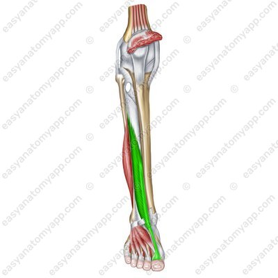

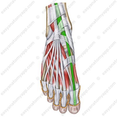
(m. extensor hallucis longus)
Origin: middle third of the anterior surface of the fibula, interosseous membrane of the leg
Insertion: base of the distal phalanx of the thumb
Function: extends the big toe
Innervation: deep fibular nerve (L4-S1)
Blood supply: anterior tibial artery
Muscles of the leg
- Gluteal region
- regio glutealis
- Femoral region
- regio femoralis
- Knee region
- regio genus
- Leg region
- regio cruralis
- Ankle region
- regio talocruralis
- Foot region
- regio pedis
- Tibialis anterior muscle
- m. tibialis anterior
- Extensor digitorum longus muscle
- m. extensor digitorum longus
- Peroneus tertius muscle
- m. peroneus tertius
- Extensor hallucis longus muscle
- m. extensor hallucis longus

