Let’s cover the structure of the diaphragm (diaphragma, m. phrenicus).
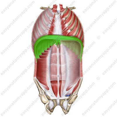
The diaphragm is a muscle-tendon structure that separates the thoracic cavity from the abdominal cavity and is classified as a respiratory muscle. The diaphragm has a domed shape, with a convex surface facing the thoracic cavity, and a concave one facing the abdominal cavity.
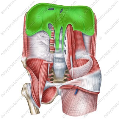
The center of the diaphragm has a connective tissue structure and is called the central tendon (centrum tendineum).
The diaphragm forms two domes:
The left dome is located at the 5th intercostal space level
The right dome is located at the 4th intercostal space level
There are three parts of the diaphragm:
The beginning of the lumbar part (pars lumbalis) is described in more detail below
Lumbar part (pars lumbalis) 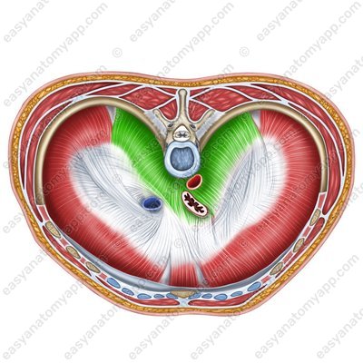
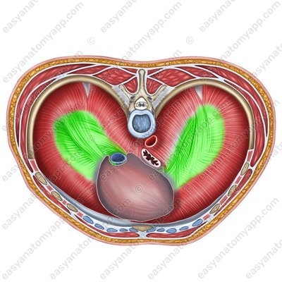
The costal part (pars costalis) starts from the inner surface of ribs 7-12
Costal part (pars costalis) 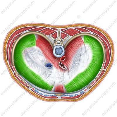
The sternal part (pars sternalis) arises from the posterior surface of the xiphoid process of the sternum
Sternal part (pars sternalis) 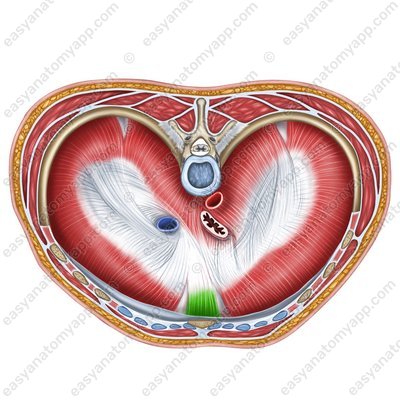
The lumbar part arises from the lumbar vertebrae (L1-L3, sometimes L4) with two cruses:
Left crus (crus sinistrum)
Left crus (crus sinistrum) 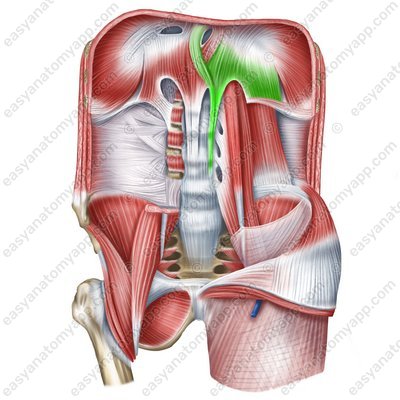
Right crus (crus dextrum)
Right crus (crus dextrum) 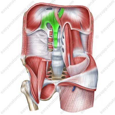
And also from the following ligaments:
Lateral arcuate ligament (lig. arcuatum laterale)
Lateral arcuate ligament (lig. arcuatum laterale) 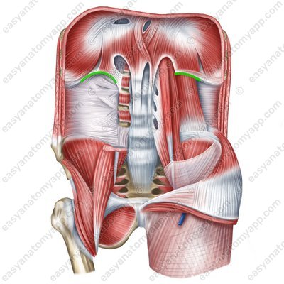
Medial arcuate ligament (lig. arcuatum mediale)
Medial arcuate ligament (lig. arcuatum mediale) 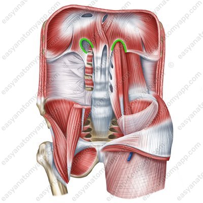
Median arcuate ligament (lig. arcuatum medianum)
Median arcuate ligament (lig. arcuatum medianum) 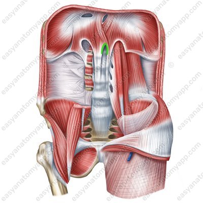
Anterior longitudinal ligament (lig. longitudinale anterius)
Anterior longitudinal ligament (lig. longitudinale anterius) 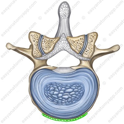
The ligaments are intertwined in various ways, as a result of which several hiatuses are formed:
The aortic hiatus (hiatus aorticus), in which the aorta and thoracic lymph duct pass. Here, the median arcuate ligament protects the aorta from compression during the contraction of the diaphragm.
Aortic hiatus (hiatus aorticus) 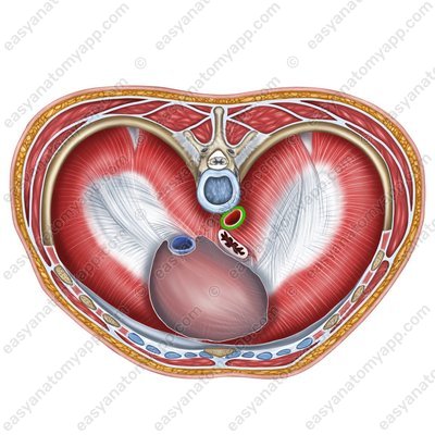
The esophageal hiatus (hiatus oesophageus) is where the esophagus and the vagus nerves pass. At this site, the diaphragm performs the function of the esophageal sphincter of sorts. However, this site is also a so-called “weak spot”, where hernias may form.
Oesophageal hiatus (hiatus oesophageus) 
Also, the sympathetic trunk (truncus sympathicus), great and lesser splanchnic nerves (N. splanchnicus major et n. splanchnicus minor), azygos (v. azygos) and hemi-azygos veins (v. hemiazygos) pass between the cruses of the diaphragm.
There are two structures in the area of the central tendon:
Cardiac impression (impressio cordis)
Opening of inferior vena cava (foramen venae cavae inferioris)
Caval opening (foramen venae cavae inferioris) 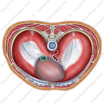
On the side of the abdominal cavity, the diaphragm is covered by the intra-abdominal fascia and parietal peritoneum, and on the side of the thoracic cavity, it is covered by the endothoracic fascia and parietal diaphragmatic pleura.
Between the parts of the diaphragm, there are regions devoid of muscle fibers. They are called triangles of the diaphragm. These are also “weak spots”, where hernias often form.
Lumbocostal triangle / Bochdalek’s triangle (trigonum lumbocostale)
Lumbocostal triangle (trigonum lumbocostale) 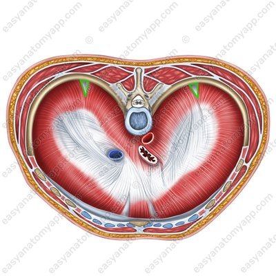
Sternocostal triangle / on the right: Morgagni’s triangle / on the left: Larrey’s triangle (trigonum sternocostale)
The diaphragm is the primary respiratory muscle. During inhalation, it flattens, due to which the volume of the thoracic cavity increases. During exhalation, on the contrary, it becomes convex, reducing the volume of the thoracic cavity.
In addition, the diaphragm participates in increasing intra-abdominal pressure during various physiological processes, including blood circulation, defecation, urination, childbirth, lymph drainage.
Innervation: phrenic nerve (C3-C5), sensory innervation — intercostal nerves 6-11
Blood supply: musculophrenic artery, pericardiacophrenic artery, superior and inferior phrenic arteries, posterior intercostal arteries.
Diaphragm
- Diaphragm
- diaphragma, m. phrenicus
- Central tendon
- centrum tendineum
- Lumbar part
- pars lumbalis
- Costal part
- pars costalis
- Sternal part
- pars sternalis
- Left crus
- crus sinistrum
- Right crus
- crus dextrum
- Lateral arcuate ligament
- lig. arcuatum laterale
- Medial arcuate ligament
- lig. arcuatum mediale
- Median arcuate ligament
- lig. arcuatum medianum
- Anterior longitudinal ligament
- lig. longitudinale anterius
- Aortic hiatus
- hiatus aorticus
- Oesophageal hiatus
- hiatus oesophageus
- Cardiac impression
- impressio cordis
- Caval opening
- foramen venae cavae inferioris
- Lumbocostal triangle
- trigonum lumbocostale
- Sternocostal triangle
- trigonum sternocostale

