In this note, we will consider the anatomy of the esophagus (oesophagus).
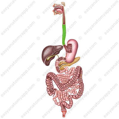

This is an unpaired fibromuscular organ that provides the passage of food from the pharynx to the stomach.
It is located in the neck cavity, as well as thoracic and abdominal cavities.
It arises at the level of the superior border of the 7th cervical vertebra and ends at the level between the 11th and 12th thoracic vertebrae.
There are 3 parts of the esophagus:
- Сervical part (pars cervicalis)
- Thoracic part (pars thoracica)
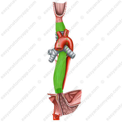
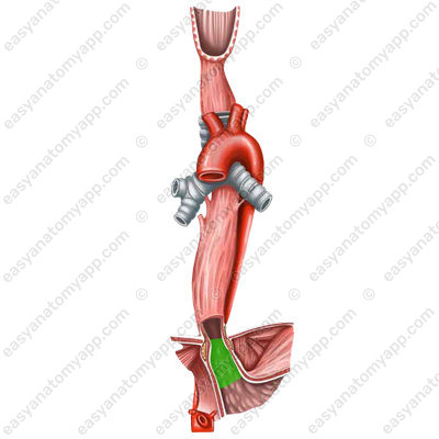
- Abdominal part (pars abdominalis)
The cervical part is located posteriorly to the trachea and anteriorly to the vertebral column. On the lateral regions of the neck, there is a neurovascular bundle of the neck, consisting of the common carotid artery, internal jugular vein, and vagus nerve.
At the level of the 3rd thoracic vertebra anteriorly to the esophagus, there is the aortic arch, and at the level of the 9th thoracic vertebra, the thoracic aorta lies posteriorly to the esophagus. In other words, the esophagus wraps around the aorta spirally.
The esophagus has three anatomical constrictions:
- Pharyngeal constriction, which is located at the level of the cricoid cartilage of the larynx (C6-C7)

- Bronchial constriction, which is located at the site where the esophagus crosses the left main bronchus (Th4-Th5)

- Diaphragmatic constriction, which is located at the level of the esophageal hiatus of the diaphragm (Th10-Th11)

In addition to the anatomical constrictions, there are also functional constrictions of the esophagus:
- Aortic constriction, which is formed due to the adherence of the aortic arch (Th3)
- Cardiac constriction, which is formed due to the tension of the cardiac sphincter (at the level where the esophagus passes into the stomach, Th11-Th12)
The wall of the esophagus consists of several layers
1. Mucous membrane (tunica mucosa), which is lined with a nonkeratinizing multilayered squamous epithelium, which, when the esophagus passes into the stomach, is replaced by a single-layered simple columnar epithelium of the mucous membrane of the stomach.
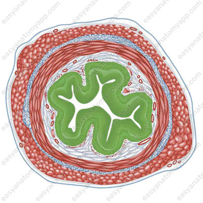
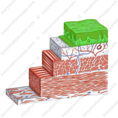
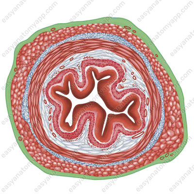
2. Submucosa (tela submucosa), is well-developed. Because of it, the mucous membrane forms longitudinal folds, therefore the lumen of the esophagus has a stellate shape on the cross-section. In the submucosa, there are numerous glands of the esophagus.

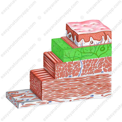
3. Muscular layer (tunica muscularis), is formed in the superior third by striated muscle fibers, which are gradually replaced by smooth myocytes in the middle part, and in the inferior part, this layer consists entirely of smooth myocytes.
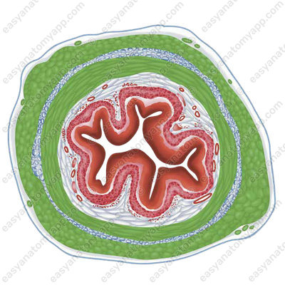
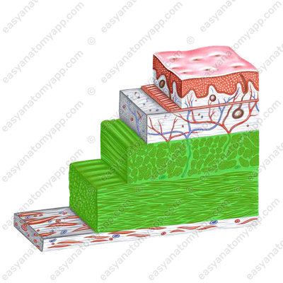
The muscle fibers are arranged in two layers.
- Internal circular layer (stratum circulare)
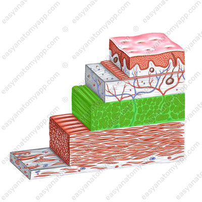
- External longitudinal layer (stratum longitudinale)
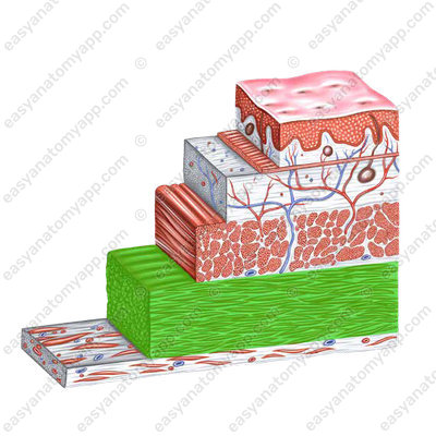
4. Adventitia (tunica adventitia) is formed by loose connective tissue. It covers the cervical and thoracic parts of the esophagus.
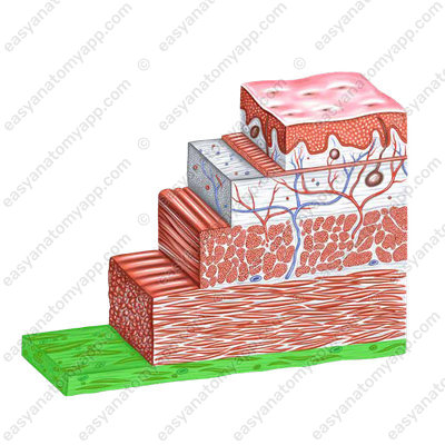
The abdominal part of the esophagus is completely wrapped by the visceral peritoneum.
Blood supply
The cervical part is supplied with blood by esophageal branches (rr. esophageales) from the inferior thyroid artery (a. thyroidea inferior)
The thoracic part is supplied with blood by esophageal branches (rr. esophageales) from the thoracic aorta (pars thoracica aortae)
The abdominal part is supplied with blood by esophageal branches (rr. esophageales) from the inferior phrenic arteries (aa. phrenicae inferiores) and the left gastric artery (a. gastrica sinistra)
Venous drainage
Through the eponymous veins:
from the cervical part, the blood drains to the inferior thyroid vein (v. thyroidea inferior)
from the thoracic part, the blood drains to the azygos and hemi-azygos veins (vv. azygos et hemiazygos)
from the abdominal part, the blood drains to the left gastric vein (v. gastrica sinistra)
Lymphatic drainage
- Internal jugular lymph nodes (nodi lymphatici jugulares interni)
- Prevertebral lymph nodes (nodi lymphatici prevertebrales)
- Posterior mediastinal lymph nodes (nodi lymphatici mediastinalis posteriores)
- Left gastric lymph nodes (nodi lymphatici gastricae sinistrum)
Innervation
Esophageal branches of the right and left vagus nerves (n. vagus)
Anatomy of the esophagus
- Esophagus
- oesophagus
- Cervical part
- pars cervicalis
- Thoracic part
- pars thoracica
- Abdominal part
- pars abdominalis
- Mucous membrane
- tunica mucosa
- Submucosa
- tela submucosa
- Muscular layer
- tunica muscularis
- Circular layer
- stratum circulare
- Longitudinal layer
- stratum longitudinale
- Adventitia
- tunica adventitia

