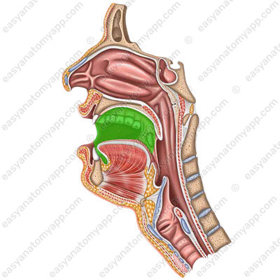
In this note, we will consider the anatomy of the initial part of the digestive tract, called the oral cavity (cavitas oris).
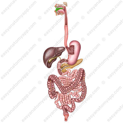

Due to the presence of organs such as salivary glands, teeth, and tongue in the oral cavity and adjacent tissues, the food may be ground, mixed and moistened with saliva.
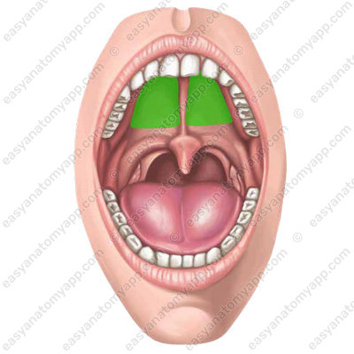
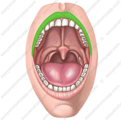
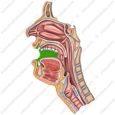
The oral cavity has the following borders:
- The hard and soft palate are the superior border
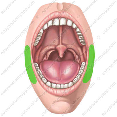
- The cheeks are the lateral borders
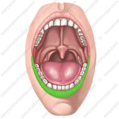
- The muscles (the so-called “floor of the oral cavity”) are the inferior border
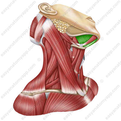
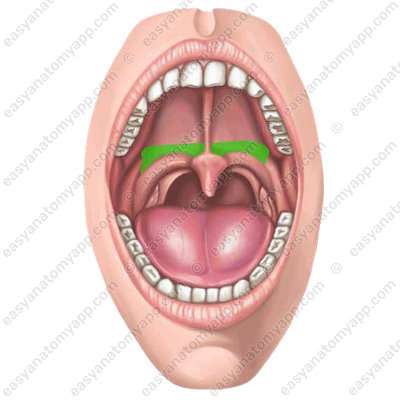
- Inferiorly, the oral cavity is connected to the pharynx via the wide opening called the fauces (fauces)

Anteriorly, the oral cavity is delimited by the lips. These are the upper lip (labium superius) and the lower lip (labium inferius)
These are the folds made of skin and muscles, and the buccolabial muscular group lies at the base of these folds. There are three sections of the lip:
- Mucous section
- Transitional section (or the so-called vermilion border)
- Cutaneous section
Laterally, the lips are fused by a labial commissure (comissura labiorum)

The oral cavity is divided into two parts:
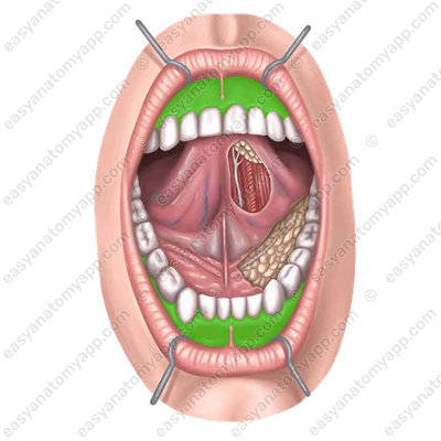
- The oral vestibule (vestibulum oris)
- The oral cavity proper (cavitas oris propria)
They are separated from each other by the alveolar processes of the jaws, gingivae (or gums), and teeth.
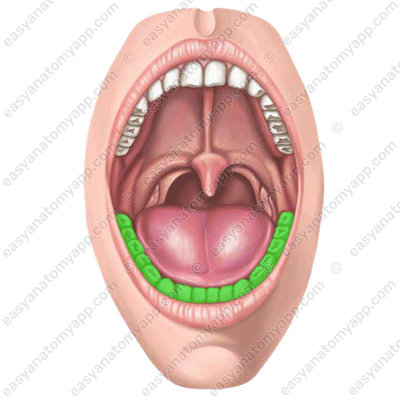
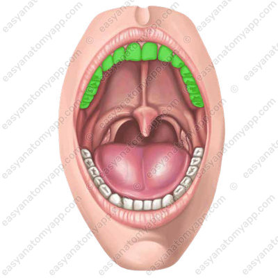
In turn, the gums are the alveolar processes of the jaws covered with soft tissues and mucous membrane.
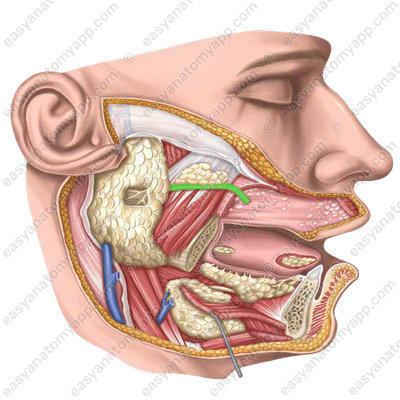
In the oral vestibule, at the level of the second upper molar tooth, the excretory duct of the parotid salivary gland (ductus parotideus) opens in the mucous membrane of the cheek.
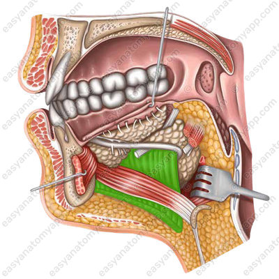
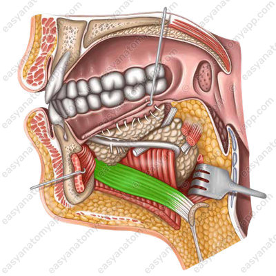
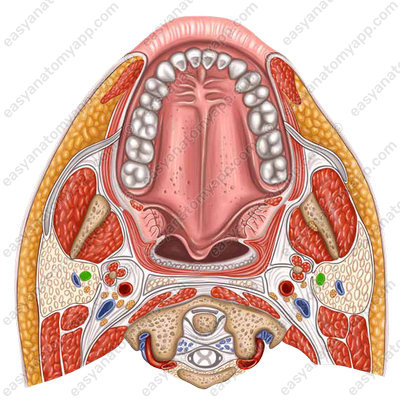
The muscular diaphragm of the oral cavity (or the so-called floor of the oral cavity) is represented by mylohyoid and geniohyoid muscles, which are covered by a mucous membrane from the inside.
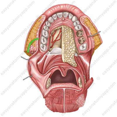
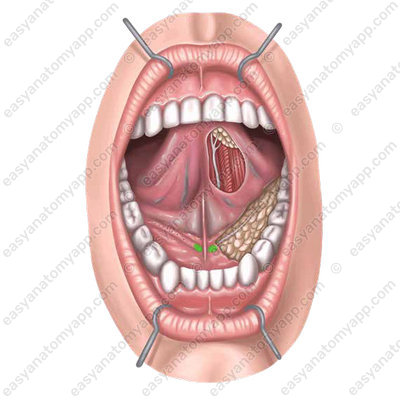
At the bottom of the oral cavity at the margin of the tongue, there are paired sublingual folds (plural – plicae sublinguales; singular – plica sublingualis).

The anterior closed part of these folds has an elevation called the sublingual caruncle (caruncula sublingualis), on which the excretory ducts of the submandibular and sublingual salivary glands open.
The cheek (bucca) is the lateral wall of the oral cavity, it has the buccinator muscle as its base.

Between the skin and the muscle, there is the buccal fat pad (corpus adiposum buccae).
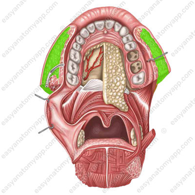
It is also called Bichat’s fat pad, and it is especially well-developed in children.
Blood supply
The branches of the external carotid artery (a. carotis externa) supply the oral cavity with arterial blood.
Venous drainage
The external jugular vein (v. jugularis externa) drains venous blood from the oral cavity.

Innervation
It is innervated by the trigeminal, facial, glossopharyngeal, vagus, and sublingual nerves.
Anatomy of the oral cavity
- digestive system
- systema digestorium
- lip
- labium
- oral cavity
- cavitas oris
- cheeks
- buccae
- tongue
- lingua
- pharynx
- pharynx
- oral cavity proper
- cavitas oris
- oral vestibule
- vestibulum oris
- oral cavity proper
- cavitas oris propria
- parotid duct
- ductus parotideus
- fauces
- fauces
- sublingual folds
- plicae sublinguales
- sublingual caruncle
- caruncula sublingualis
- gingivae (gums)
- gingivae
- upper lip
- labium superius
- lower lip
- labium inferius

