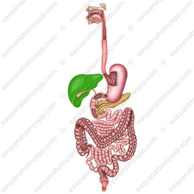
The liver (hepar) is the largest gland in the human body.
Function
It performs a number of important functions in the human body:
- Neutralization of various foreign substances
- Neutralization and removal of excess hormones, mediators, vitamins, as well as toxic intermediates and end products of metabolism from the body
- Providing the energy needs of the body with glucose and converting various energy sources (free fatty acids, amino acids, glycerin, lactic acid, etc.) into glucose
- Replenishment and storage of rapidly mobilized energy reserves in the form of glycogen and regulation of carbohydrate metabolism
- Replenishment and storage of depots of some vitamins (especially large reserves of fat-soluble vitamins A, D, and water-soluble vitamin B12 in the liver), as well as depots of cations of a number of trace substances, including metals, in particular, iron, copper and cobalt cations. Also, the liver is directly involved in the metabolism of vitamins A, B, C, D, E, K, PP, and folic acid
- Participation in the processes of hematopoiesis (only in the fetus), in particular, the synthesisof many plasma proteins, including albumins, alpha and beta globulins, transport proteins for various hormones and vitamins, proteins of the coagulation and anticoagulation systems of the blood and many others; the liver is one of the important organs of hematopoiesis in prenatal development
- Synthesis of cholesterol and its esters, lipids and phospholipids, lipoproteins, and regulation of lipid metabolism
- Synthesis of bile acids and bilirubin, production and secretion of bile
- It also serves as a depot for a fairly significant volume of blood, which can be discharged into the general circulation in case of blood loss or shock due to dilation of the vessels supplying the liver
- Synthesis of hormones (for example, insulin-like growth factors).
- Participation in pigment metabolism (the pigment called bilirubin is formed in the liver as a result of the destruction of red blood cells and is excreted with bile)
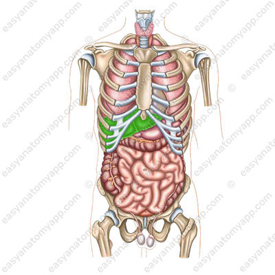
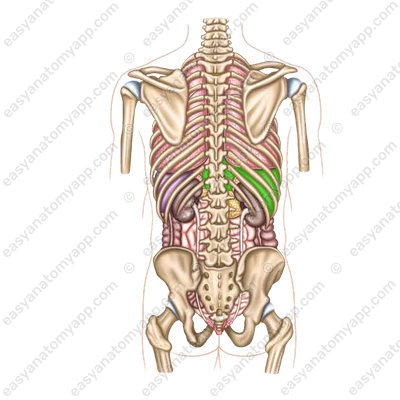
Location & Borders
It is located mainly in the right subcostal region and in the epigastric region. A small part of the organ is located in the left epigastric region.
The superior border of the liver passes as follows:
- Along the right midclavicular line, it runs at the level of the 5th rib.
- Along the anterior median line, it runs at the level of the base of the xiphoid process.
- Along the parasternal line it runs at the level of the cartilage of the 6th rib.
The inferior border of the liver passes as follows:
- On the right, it coincides with the inferior border of the costal arch
- Then it comes out from under the ribs at the junction of the 8th and 9th ribs on the right
- Then it is directed to the left and upward through the apex of the xiphoid process to the level of the cartilages of the 8th and 7th left ribs, at the place of their attachment to the sternum.

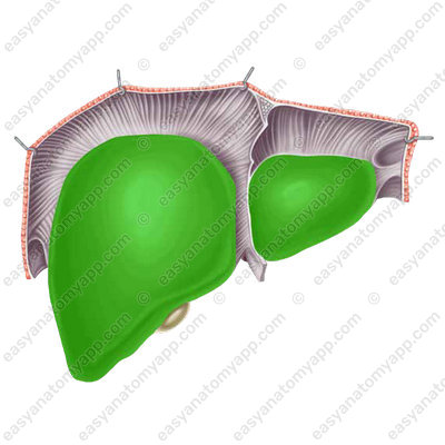
The organ has two surfaces:
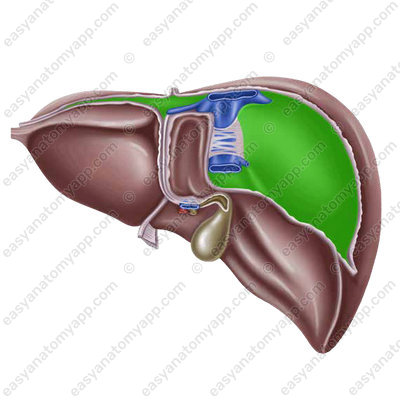
1. The diaphragmatic surface (facies diaphragmatica)
2. The visceral surface (facies visceralis)

Also, the liver has two margins:
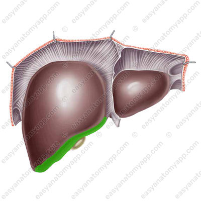
1. The inferior margin (margo inferior)
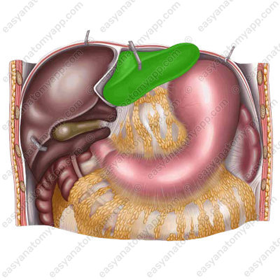
2. The posterior margin (margo posterior), to which the esophagus adheres
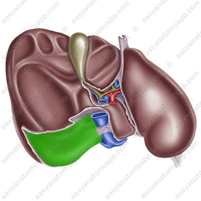
The organ consists of two main lobes:
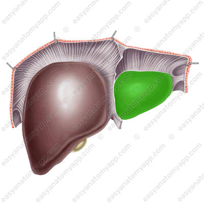
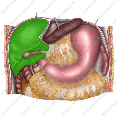
1. The left lobe (lobus sinister), to which the stomach adheres
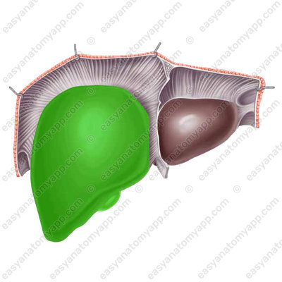
2. The right lobe (lobus dexter), to which the colon, the right kidney, the right adrenal gland, and the duodenum adhere.
The right lobe, in turn, has secondary lobules in its structure:
- The quadrate lobe (lobus quadratus), to which the stomach also adheres
- The caudate lobe (lobus caudatus)
The caudate lobe has the following processes:
- The papillary process (processus papillaris)
- The caudate process (processus caudatus)
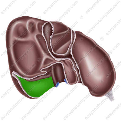
If we take a look at the visceral (inferior) surface of the liver, we will see the following grooves:
- The left longitudinal sulcus (sulcus longitudinalis sinister), which is filled in front with the round ligament of the liver (this is the obliterated umbilical vein), and in the back it is filled with the ligamentum venosum (the obliterated venous duct).
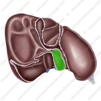
- The right longitudinal sulcus (sulcus longitudinalis dexter), which is filled in front with the gallbladder and in the back with the inferior vena cava.
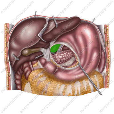
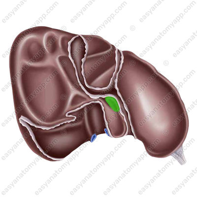
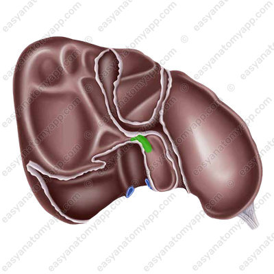
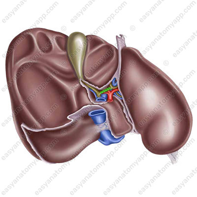
- The transverse sulcus (sulcus transversus), where the vessels and nerves are located. It is also called the porta hepatis (porta hepatis).
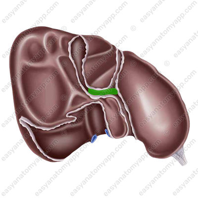
The porta hepatis includes:
- Portal vein
- Proper hepatic artery
- Nerves
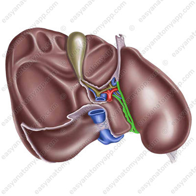
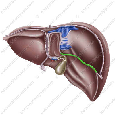
The following structures exit from the porta hepatis:
- Common hepatic duct (ductus hepaticus communis)
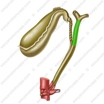
- Lymphatic vessels
The liver has 9 ligaments, which are divided into 3 groups. Let’s consider them.
Ligaments of the diaphragmatic surface:
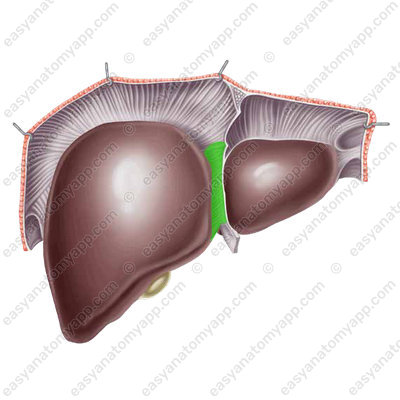
1. Falciform ligament of the liver (lig. falciformis hepatis)
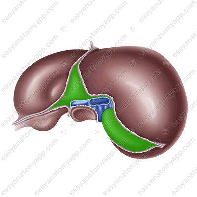
2. The coronary ligament of the liver (lig. coronarium hepatis)

3. The left triangular ligament of the liver (lig. triangularis sinister)

4. The right triangular ligament of the liver (lig. triangularis dexter)

Ligaments of the visceral surface of the liver:
1. The round ligament of the liver (lig. teres hepatis)

2. The ligamentum venosum (lig. venosum)
Ligaments, which insert into neighboring organs:
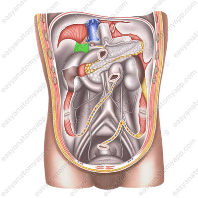
1. The hepatorenal ligament (lig. hepatorenale)
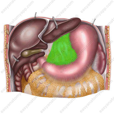
2. The hepatogastric ligament (lig. hepatogastricum)

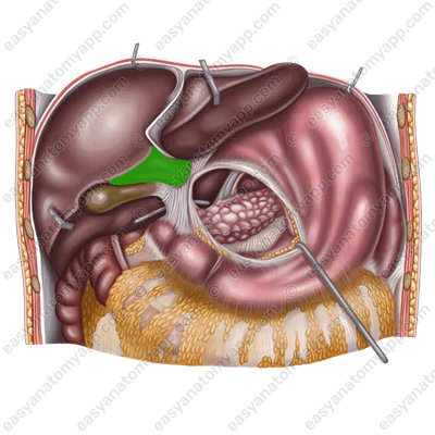
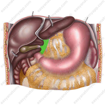
3. The hepatoduodenal ligament (lig. hepatoduodenale)
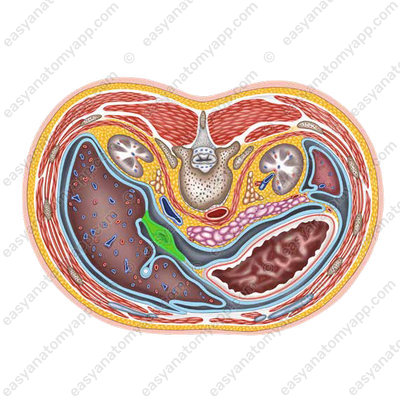
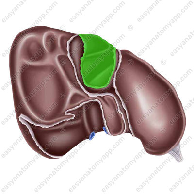
The liver is located mesoperitoneally. In the superior part, the liver fuses with the diaphragm, the place of fusion is called the bare area (area nuda). The peritoneum itself is tightly fused with the liver by a fibrous capsule (tunica fibrosa) and forms the capsule of the liver (Glisson’s capsule).
The liver is held in its place by a number of anatomical structures, such as the ligaments and vessels already mentioned, as well as by intra-abdominal pressure.
Segments
The Couinaud classification of liver segments is the most widely used classification system in describing the anatomy of the liver. According to this classification, the liver is divided into eight independent functional units called segments. The segments are numbered with numerals from I to VIII.
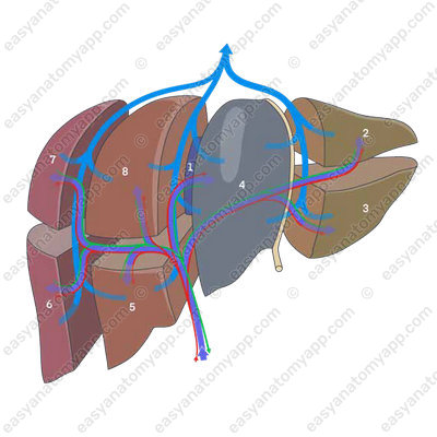
The division into segments is based on the fact that each segment has its own dual blood supply, as well as bile and lymph outflow tracts. Each segment is wedge-shaped with its apex directed to the hilum of the liver. In the region of the apex, the segment includes the segmental branch of the portal vein, hepatic artery, and bile duct. The borders of the segments are the hepatic veins, each of which drains two or more adjacent segments.
Microscopic anatomy
Let’s briefly consider the main features of the microscopic structure of the liver.
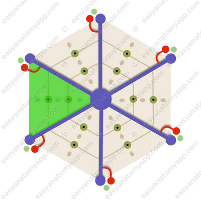
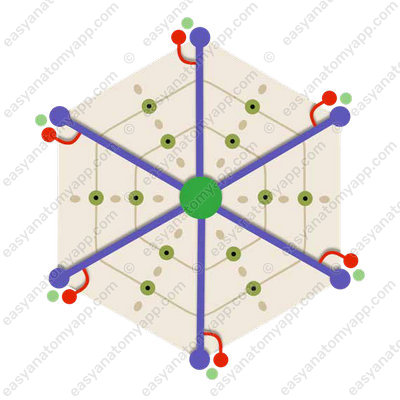
The structural and functional unit of the liver is the hepatic lobule (lobulus hepatis)
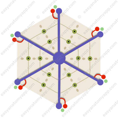
This is a section of the liver parenchyma separated by a thin layer of connective tissue, shaped like a hexagonal prism, and consisting of hepatic rods.
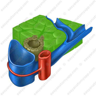
Each rod consists of hepatic cells called hepatocytes.

Bile ducts are located between these cells in each rod.
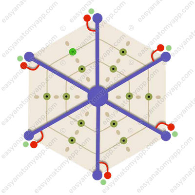
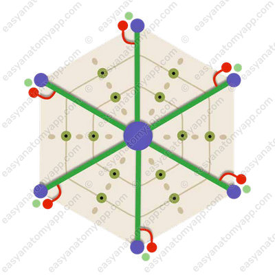
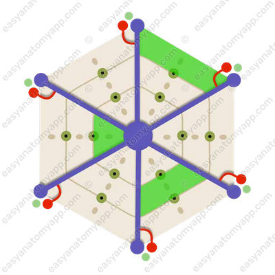
And between the adjacent rods, there are circulatory sinusoidal capillaries that converge in the center of the lobule to its central vein (v. centralis).
The hepatic lobule is penetrated by the interlobular veins from the system of the portal vein and the interlobular arteries from the system of the hepatic artery.
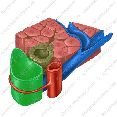
They merge into the already mentioned sinusoidal capillary.
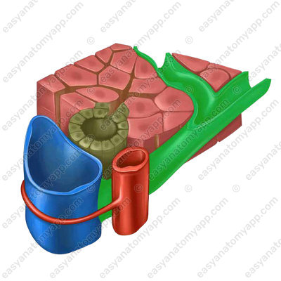
The blood drains from the capillary network into the central vein, by which the blood flows into the interlobular collecting veins. The latter then form hepatic veins, which end by the inferior vena cava.
This double type of blood circulation is called the rete mirabile (rete mirabile hepatis). The blood coming from the portal vein has already passed through the capillaries into the gastrointestinal tract, but in the liver, it again enters the capillary vessels.

Blood supply
- The proper hepatic artery (a. hepatic propria) contains arterial blood, about 30% of the blood supplied to the liver.
- The portal vein of the liver (v. portae) contains venous blood, about 70% of the blood supplied to the liver.
Venous drainage
- Hepatic veins (vv. hepaticae)
Lymph drainage
- Hepatic lymph nodes (nodi lymphatici hepatici)
- Coeliac lymph nodes (nodi lymphatici coeliaci)
- Right lumbar lymph nodes (nodi lymphatici lumbales dextri)
- Superior diaphragmatic lymph nodes (nodi lymphatici phrenici superiores)
- Inferior diaphragmatic lymph nodes (nodi lymphatici inferiores)
Innervation
Nerve fibers form the hepatic plexus (plexus hepaticus)
- Afferent innervation is provided by the anterior branches of the inferior thoracic spinal nerves and the vagus nerve.
- Sympathetic innervation is carried out by the hepatic plexus.
- Parasympathetic innervation is provided by the vagus nerve.
Anatomy of the liver
- Diaphragmatic surface
- facies diaphragmatica
- Visceral surface
- facies visceralis
- Inferior border
- margo inferior
- Posterior border
- margo posterior
- Left lobe
- lobus sinister
- Right lobe
- lobus dexter
- Quadrate lobe
- lobus quadratus
- Caudate lobe
- lobus caudatus
- Papillary process
- processus papillaris
- Caudate process
- processus caudatus
- Left paracolic gutter
- sulcus longitudinalis sinister
- Right paracolic gutter
- sulcus longitudinalis dexter
- Transverse gutter
- sulcus transversus
- Porta hepatis
- porta hepatis
- Common hepatic duct
- ductus hepaticus communis
- Falciform ligament of the liver
- lig. falciformis hepatis
- Coronary ligament of the liver
- lig. coronarium hepatis
- Left triangular ligament of the liver
- lig. triangularis sinister
- Right triangular ligament of the liver
- lig. triangularis dexter
- Round ligament of the liver
- lig. teres hepatis
- Ligamentum venosum
- lig. venosum
- Hepatorenal ligament
- lig. hepatorenale
- Hepatogastric ligament
- lig. hepatogastricum
- Hepatoduodenal ligament
- lig. hepatoduodenale
- Bare area
- area nuda
- Fibrous capsule
- tunica fibrosa
- Proper hepatic artery
- a. hepatica propria
- Portal vein of the liver
- v. portae
- Hepatic veins
- vv. hepaticae
- Hepatic lymph nodes
- nodi lymphatici hepatici
- Coeliac lymph nodes
- nodi lymphatici coeliaci
- Right lumbar lymph nodes
- nodi lymphatici lumbales dextri
- Superior diaphragmatic lymph nodes
- nodi lymphatici phrenici superiores
- Inferior diaphragmatic lymph nodes
- nodi lymphatici inferiores
- Hepatic plexus
- plexus hepaticus
- Hepatic lobule
- lobulus hepatis
- Central vein
- v. centralis
- Rete mirabile of the liver
- rete mirabile hepatis

