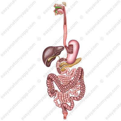
Teeth (dentes) are the organs of the digestive system, which are located in the alveoli of the jaws and take part in the mechanical processing of food and articulation of speech.
The roots are rigidly fused with the periosteum of the alveoli, forming a synarthrosis, which is calledgomphosis or dento-alveolar syndesmosis.
An adult has 32 permanent teeth, and a child has 20 deciduous teeth.
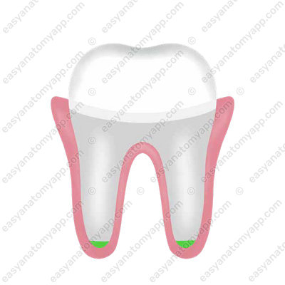
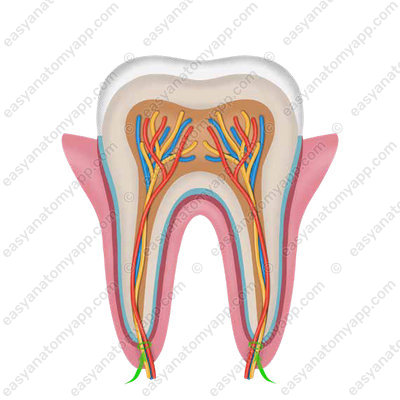
There are three main anatomical parts of the tooth:
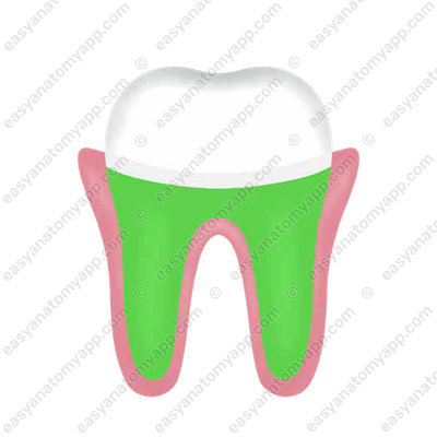
1.Crown (corona dentis), which is a thickened part of the tooth protruding from the dental alveolus covered with enamel.
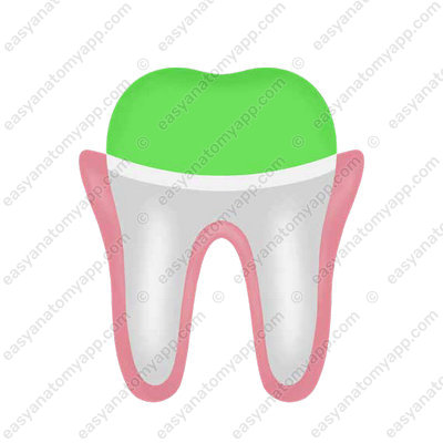
2. Neck (collum/cervix dentis), which is a narrowed part of the tooth located between the crown and the root.
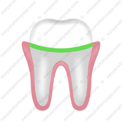
3. Root (radix dentis), which is a part of the tooth located inside the dental alveolus. The root of the tooth ends with the apex of the root (apex radicis dentis).
The crown of the tooth has the following surfaces:
1. Occlusal surface (facies occlusalis), which faces teeth of the opposite jaw.
It is present in molars and premolars. Incisors and canines end with an incisal margin (margo incisalis). Also, this surface is also called masticatory (facies masticatoria), and the margin is called masticatory (margo masticatorius) as well.
2. Vestibular surface (facies vestibularis), which faces the vestibule of the oral cavity. In the area of the frontal teeth, which touch the lips, it is also called the labial surface (facies labialis).
In the area of the posterior teeth, which adhere to the cheek, it is called the buccal surface (facies buccalis).
3. Lingual surface (facies lingualis), which faces the tongue in the oral cavity.
4. Contact surface (facies contactus), which adheres to adjacent teeth. There is the medial surface (facies medialis), which is located closer to the median line
and the lateral surface (facies lateralis), which is located closer to the edge of the alveolar arch.
In humans, as in most mammals, there are two generations of teeth: deciduous and permanent. The shape and function of teeth are closely related. In humans, there are three shapes of teeth:
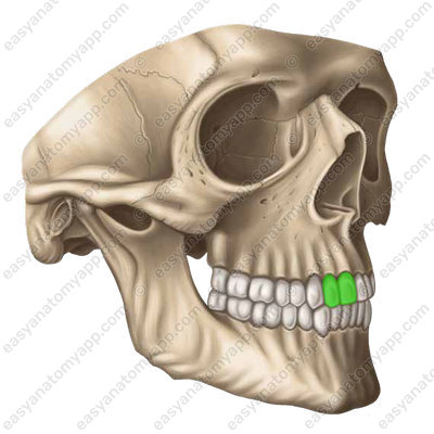
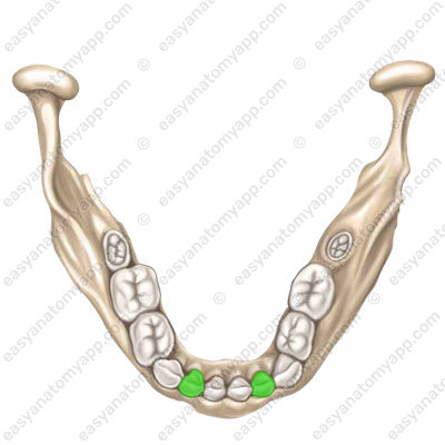
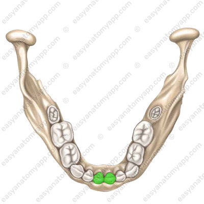
- Incisors (dentes incisive), which are used for gripping and cutting food. They have a chisel-shaped crown with a narrow incisal margin. The crown of the upper incisors is much wider than that of the lower ones. The single cone-shaped root of the incisor is narrowed laterally.
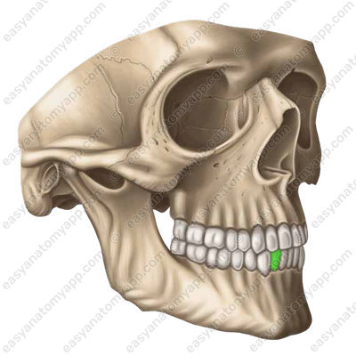

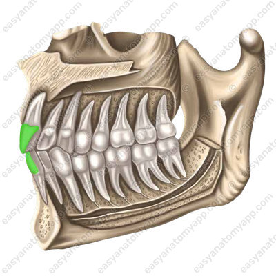
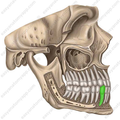
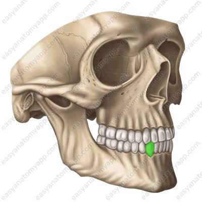
- Canines (dentes canini), which crush, and tear food. In humans, they are relatively poorly developed. They have a conical crown with a pointed cusp and a single, long, laterally compressed root, which is shorter in the lower canines than in the upper ones, and may be bifurcated at the apex of the root of the tooth. Sometimes the lower canines have a double root.

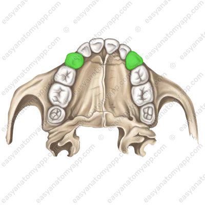

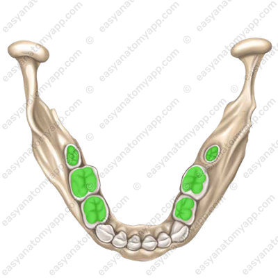
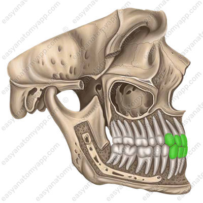
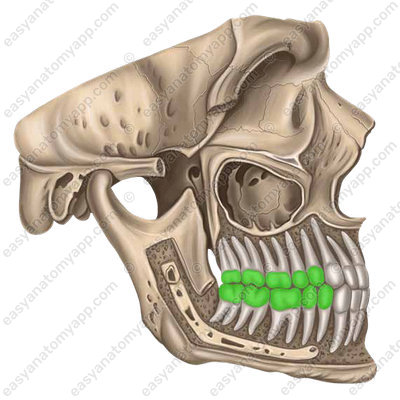
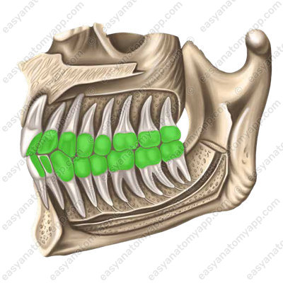
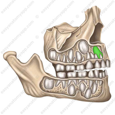
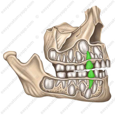
- Premolar teeth (dentes premolares) and molar teeth (dentes molares), which grind food. In addition, human teeth are involved in articulated speech. The crowns are oval, their height is much less than that of the canines. There are two conical cusps on the masticatory surface. They contribute to the grinding and crushing of food. The size of the molars reduces from front to back.

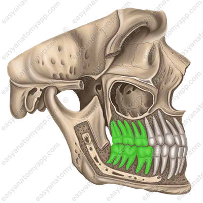
Premolar teeth usually have one root, and molar teeth have two or three roots.
Let’s briefly consider the main features of the histological structure of teeth. The tooth consists mainly of dentine (dentinum)

In the root area, it is covered with cement (cementum);
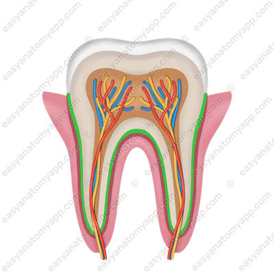
And in the area of the crown, it is covered with enamel (enamelum).

Enamel (enamelum) consists mainly of inorganic salts, such as phosphoric acid, fluoride, calcium carbonate, etc.
Dentine (dentinum), about a third of which are organic substances (mainly collagen), and the rest are inorganic substances (calcium phosphate, magnesium, calcium fluoride admixture).
Cement (cementum) is similar to bone tissue in its composition.
Inside the tooth there is a cavity filled with dental pulp (pulpa dentis), which is rich in blood vessels and nerves.
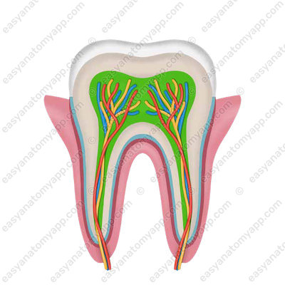
The cavity continues into the root canal of the tooth (canalis radicis dentis).
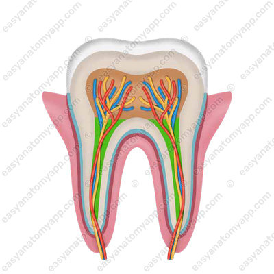
It ends with an apical foramen (foramen apicis dentis).
The roots of the teeth are tightly fused with the surface of the dental alveoli by the periodontium.
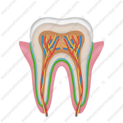
The periodontium has bundles of connective tissue fibers that insert, on the one hand, into the bone of the alveoli (Sharpey’s fibers), and, on the other, into the cement of the root of the tooth. In the area of the neck of the tooth, it forms a circular ligament.
It is important to note that the concept of the periodontium is also distinguished, which includes gums, periodontal, cement, and alveolar processes.
Human teeth are arranged symmetrically in the form of two dentitions: upper and lower. Teeth are commonly referred to according to the “dental formula”.
In clinical practice, FDI-ISO system is the most commonly used tooth numbering system. The upper row corresponds to the maxilla, while the lower raw corresponds to the mandible. The left side of the image corresponds to the teeth of the RIGHT side, and the right side of the image corresponds to the teeth of the LEFT side.
| FDI World Dental Federation notation | |||||||||||||||
|---|---|---|---|---|---|---|---|---|---|---|---|---|---|---|---|
| Permanent teeth | |||||||||||||||
| 1.8 | 1.7 | 1.6 | 1.5 | 1.4 | 1.3 | 1.2 | 1.1 | 2.1 | 2.2 | 2.3 | 2.4 | 2.5 | 2.6 | 2.7 | 2.8 |
| 4.8 | 4.7 | 4.6 | 4.5 | 4.4 | 4.3 | 4.2 | 4.1 | 3.1 | 3.2 | 3.3 | 3.4 | 3.5 | 3.6 | 3.7 | 3.8 |
The deciduous teeth in children are different from the permanent teeth in adults. In the pediatric dental formula, the deciduous teeth are designated as follows:
| FDI World Dental Federation notation | |||||||||
|---|---|---|---|---|---|---|---|---|---|
| Deciduous teeth (baby teeth) | |||||||||
| 5.5 | 5.4 | 5.3 | 5.2 | 5.1 | 6.1 | 6.2 | 6.3 | 6.4 | 6.5 |
| 8.5 | 8.4 | 8.3 | 8.2 | 8.1 | 7.1 | 7.2 | 7.3 | 7.4 | 7.5 |
Permanent teeth (dentes permanentes), which are located between the roots of deciduous teeth before eruption. Before the eruption of a permanent tooth, the deciduous tooth falls out.
The first to erupt are the first molars, then the medial and lateral incisors, the first and second permolars and the second molars. The third molars (wisdom teeth) are the last to erupt.
Deciduous teeth (dentes decidui), which have a structure similar to the permanent ones, but their sizes are about half as small. The enamel of deciduous teeth has a matte white and bluish color. The roots of the deciduous teeth are poorly developed, and their neck is well expressed.
When the maxilla and mandible come together, the teeth of the upper and lower dentition occlude. This is called the dental occlusion. With the normal dental occlusion, the teeth of the maxilla partially overlap the teeth of the mandible due to the larger size of the superior alveolar arch and the direction of the teeth of the upper dentition outward.
Blood supply
The teeth of the maxilla are supplied with blood by the dental branches of the maxillary artery, and the teeth of the mandible are supplied by the branches of the inferior alveolar artery. Branches of the superior posterior alveolar artery also approach the posterior teeth of the maxilla, while branches of the superior anterior alveolar artery approach the anterior teeth of the maxilla. The dental arterial branches enter the pulp cavity through the apical foramen of the root of the tooth together with the nerves.
Venous drainage
Blood drainage occurs through the veins of the same name, which end by the facial veins.
Lymphatic drainage
Lymph flows into the submandibular, submental, and deep cervical lymph nodes.
Innervation
The teeth of the maxilla are innervated by the maxillary nerve (the second branch of the trigeminal nerve).
The teeth of the mandible are innervated by the mandibular nerve (the third branch of the trigeminal nerve).
Anatomy of the teeth
- Tooth
- dens
- Teeth
- dentes
- Crown
- corona dentis
- Neck
- collum dentis
- Root
- radix dentis
- Apex of the root
- apex radicis dentis
- Occlusal surface
- facies occlusalis
- Incisal margin
- margo incisalis
- Masticatory surface
- facies masticatoria
- Masticatory margin
- margo masticatorius
- Vestibular surface
- facies vestibularis
- Labial surface
- facies labialis
- Buccal surface
- facies buccalis
- Lingual surface
- facies lingualis
- Contact surface
- facies contactus
- Medial surface
- facies medialis
- Lateral surface
- facies lateralis
- Incisor teeth
- dentes incisive
- Canine teeth
- dentes canini
- Premolar
- dentes premolares
- and molar teeth
- et molares
- Dental pulp
- pulpa dentis
- Enamel
- enamelum
- Dentine
- dentinum
- Cementum
- cementum
- Permanent teeth
- dentes permanentes
- Deciduous teeth
- dentes decidui

