In this note, we will consider the anatomy of the pharynx (pharynx).
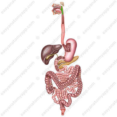
This is an unpaired organ that carries food from the oral cavity into the esophagus, as well as air from the nasal cavity into the larynx.
It is a funnel-shaped muscular canal with a length of 11-12 cm.
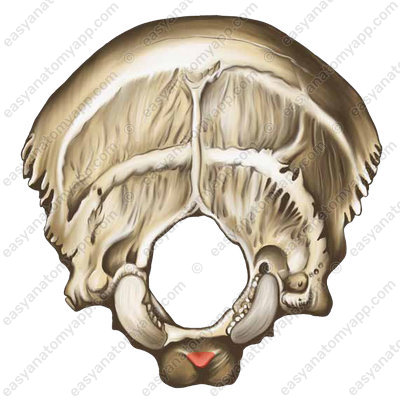
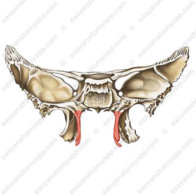
The superior part of the pharynx is called the fornix (fornix pharyngis) and is attached to the pharyngeal tubercle of the occipital bone (tuberculum pharyngeum).

The lateral part of the pharynx attaches to the petrous part of the temporal bone
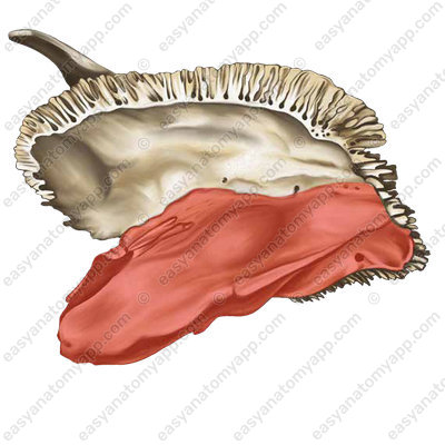
and the medial lamina of the pterygoid process.
The inferior part of the pharynx reaches the inferior border of the sixth cervical vertebra, where it passes into the esophagus.
There are the following structures located anteriorly to the pharynx:
- Nasal cavity
- Oral cavity proper
- Larynx
Deep muscles of the neck are located behind the pharynx. In addition, between the posterior surface of the pharynx and the lamina of the cervical fascia, there is a retropharyngeal space filled with loose connective tissue, in which the retropharyngeal lymph nodes are located. The pharynx is mobile due to the loose connective tissue.
Laterally to the pharynx, there is the neurovascular bundle of the neck, which contains the carotid artery, internal jugular vein, and vagus nerve
The pharynx consists of three parts.
- Nasal part (pars nasalis), which is also called the nasopharynx.
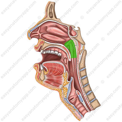
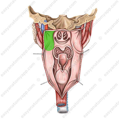
It communicates with the nasal cavity via the choanae (choanae).
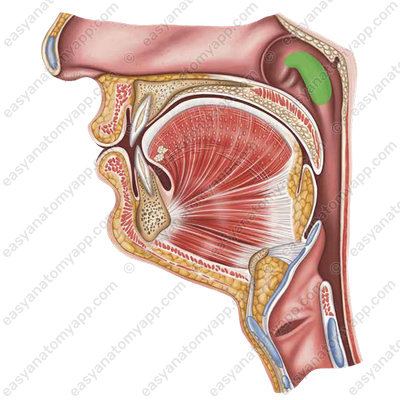
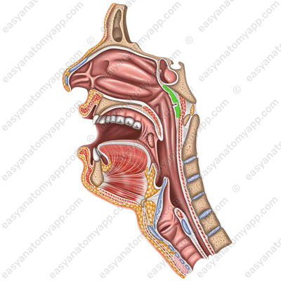
And via the pharyngeal opening of the auditory tube (ostium pharyngeum tubae auditivae) it communicates with the tympanic cavity.

The pharyngeal opening of the auditory tube is delimited by the torus tubarius (torus tubarius).

which continues downward into the salpingopharyngeal fold (plica salpingopharyngea)

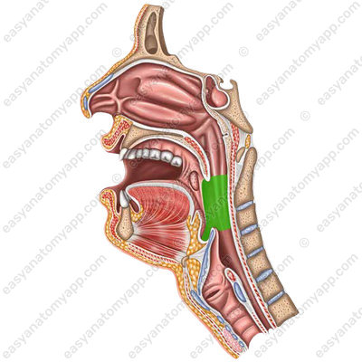
- Oral part (pars oralis), which is also called the oropharynx. It communicates with the oral cavity via the fauces (fauces).
- Laryngeal part (pars laryngea), which is the so-called laryngopharynx.
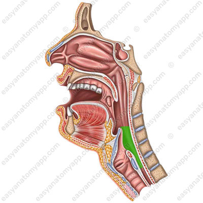
This is where the laryngeal inlet (aditus laryngis) is located;
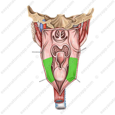
there is also the piriform recess (recessus piriformis).

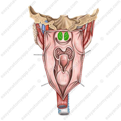
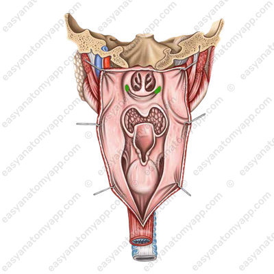
At the pharyngeal inlet, there is a complex of lymphoid formations that make up the pharyngeal lymphoid ring (or Waldeyer’s Ring), which plays an important role in the functioning of the immune system.
It consists of:
Lingual tonsil (tonsilla lingualis)
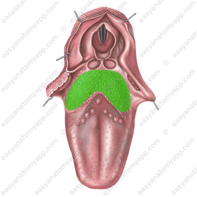
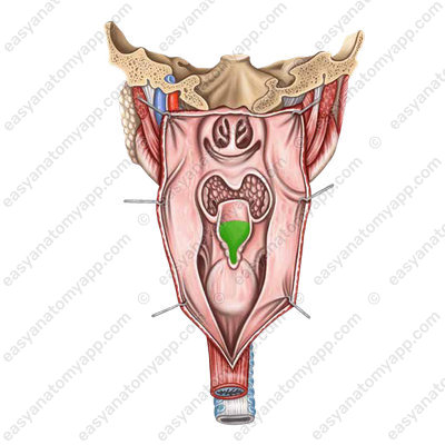
Pharyngeal tonsil (tonsilla pharyngea)
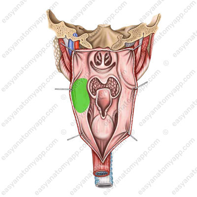
Palatine tonsil (tonsilla palatina), which is paired
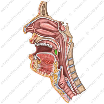
Tubal tonsil (tonsilla tubaria), which is paired and located in the pharyngeal opening of the auditory tube.
Pharyngeal muscles
The pharyngeal muscles are divided into two types:
- Longitudinal muscles
- Circular muscles (or constrictor muscles)
The longitudinal muscles include:
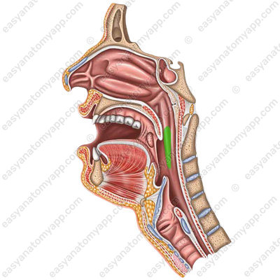
Stylopharyngeal muscle (m. stylopharyngeus), which arises from the styloid process of the temporal bone and inserts into the lateral wall of the pharynx.

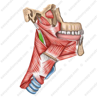
Function: raises the pharynx.
Innervation: glossopharyngeal nerve (n. glossopharyngeus) , which is the 9th pair of cranial nerves.
Salpingopharyngeus muscle (m. salpingopharyngeus), which arises from the inferior side of the cartilage of the auditory tube and inserts into the lateral wall of the pharynx.

Function: It lifts the pharynx, lowers the soft palate, and reduces the fauces.
Innervation: glossopharyngeal nerve (n. glossopharyngeus), which is the 9th pair of cranial nerves.
The circular muscles include pharyngeal constrictors.
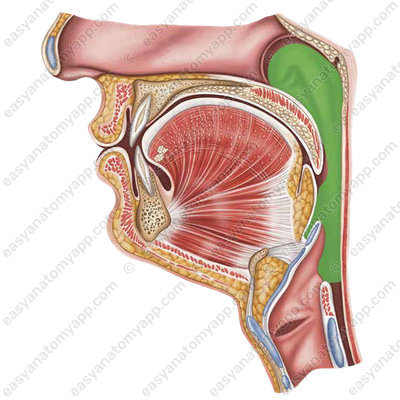
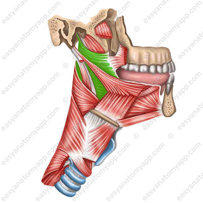
Superior constrictor of the pharynx (m. constrictor pharyngis superior) arises from the medial lamina of the pterygoid process of the sphenoid bone, the pterygomandibular raphe and the mylohyoid line of the mandible.
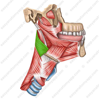
Middle constrictor of the pharynx (m. constrictor pharyngis medius) arises from the greater and lesser horns of the hyoid bone. The bundles fan out up and down, heading to the posterior surface of the pharynx, where they fuse with the muscle bundles of the opposite side.
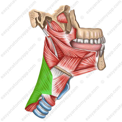
Inferior constrictor of the pharynx (m. constrictor pharyngis inferior) arises from the lateral surface of the thyroid and cricoid cartilage. On the posterior surface of the pharynx, the muscles of both sides fuse along the midline, forming a pharyngeal raphe (raphe pharyngis).

Constrictors cover each other in a tile-like manner, with the superior muscle lying deeper than the others.
Constrictor muscles contract sequentially from top to bottom during the act of swallowing, pushing a food bolus from the pharynx into the esophagus.
Innervation: pharyngeal plexus (g. pharyngis), which is formed by the fibers of the glossopharyngeal nerve (n. glossopharyngeus), this is the 9th pair of cranial nerves. The sympathetic innervation is provided by the sympathetic trunk (truncus sympathicus).
Blood supply: branches of the ascending pharyngeal artery (a. pharyngeus ascendens), pharyngeal branches of the thyrocervical trunk (rr. pharyngei) and ascending palatine artery (a. palatina ascendens).
Physiology of swallowing
In the process of swallowing, the following sequence of events occurs.
- The muscles of the soft palate contract and it rises.
- The unattached border of the soft palate is tightly pressed against the posterior wall and to the vault of the pharynx, separating the nasopharynx from the rest of the pharynx.
- Contraction of the muscles of the floor of the oral cavity leads to lifting and pulling the larynx forward. At the same time, the epiglottis closes the laryngeal inlet.
- The contraction of the styloglossus and hyoglossus muscles leads to the fact that the root of the tongue goes posteriorly and pushes the food bolus into the fauces. As a result of the contraction of the palatoglossus muscles, the food bolus is separated and pushed into the oropharynx.
- When a food bolus enters the pharyngeal cavity, the longitudinal muscles lift the pharynx upwards, as if pulling it on the food bolus.
- The pharyngeal constrictors contract sequentially from top to bottom, as a result of which the food bolus is pushed into the esophagus.
Blood supply of the pharynx
- The ascending pharyngeal artery (a. pharyngea ascendens), which arises from the facial artery (a. facialis)
- The ascending palatine artery (a. palatina ascendens), which arises from the facial artery (a. facialis)
- The descending palatine artery (a. palatina descendens), which arises from the maxillary artery (a. maxillaris)
- The superior thyroid artery (a. thyroidea superior), which arises from the external carotid artery (a. carotis externa)
- The inferior thyroid artery (a. thyroidea inferior), which arises from the thyrocervical trunk (truncus thyrocervicalis)
Venous drainage of the pharynx
Pharyngeal veins (vv. pharyngeae) drain blood into the internal jugular vein (v. jugularis interna) and into the brachiocephalic veins (vv. brachiocephalicae)
Lymphatic drainage of the pharynx
- Retropharyngeal lymph nodes (nodi lymphatici retropharyngei)
- Deep cervical lymph nodes (nodi lymphatici cervicales profundi)
Innervation of the pharynx
Afferent and parasympathetic innervation
- The vagus nerve (n. vagus)
- The glossopharyngeal nerve (n. glossopharyngeus)
Sympathetic innervation
The pharyngeal plexus (plexus pharyngeus), which arises from the superior cervical ganglion (g. cervicale superius)
Anatomy of the pharynx. Waldeyer’s tonsillar ring
- Pharynx
- pharynx
- Pharyngeal tubercle
- tuberculum pharyngeum
- Nasal part
- pars nasalis
- Pharyngeal opening of the auditory tube
- ostium pharyngeum tubaeauditivae
- Tubal tonsil
- tonsilla tubaria
- Oral part
- pars oralis
- Fauces
- fauces
- Laryngeal part
- pars laryngea
- Piriform recess
- recessus piriformis
- Vault of the pharynx
- fornix pharyngis
- Pharyngeal tonsil
- tonsilla pharyngealis
- Stylopharyngeus muscle
- m. stylopharyngeus
- Glossopharyngeal nerve
- n. glossopharyngeus
- Salpingopharyngeus muscle
- m. salpingopharyngeus
- Superior constrictor of the pharynx
- m. constrictor pharyngis superior
- Middle constrictor of the pharynx
- m. constrictor pharyngis medius
- Inferior constrictor of the pharynx
- m. constrictor pharyngis inferior

