In this note, we will consider the anatomy of the stomach (ventriculus or gaster)
Functions
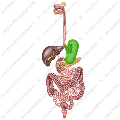
It is a muscular organ that provides accumulation, chemical and mechanical processing of food, as well as its passage into the small intestine.
Along with the chemical processing of food, the stomach performs an endocrine function (secretion of biologically active substances – histamine, gastrin, serotonin, etc.) and an absorption function (sugars, alcohol, water, salts are absorbed).
An antianemic factor is formed in the gastric mucosa, which promotes the absorption of vitamin B12 coming from food
Location and structure
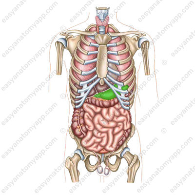
It is located in the abdominal cavity, in the left hypochondrium and in the epigastric region.
The entrance to the stomach is located at the level between the 10th and 11th thoracic vertebrae, and the exit is at the level between the 12th thoracic and 1st lumbar vertebrae, at the right border of the vertebral column.
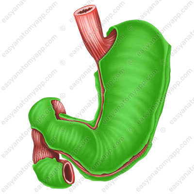
The stomach consists of the following parts.
The cardial part or cardia (pars cardiaca).
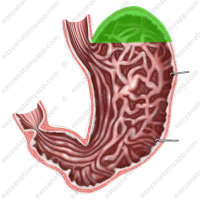
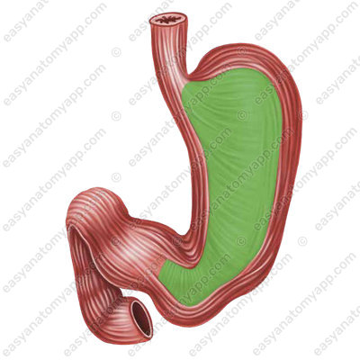
To the left of it, the stomach expands, forming the fundus (fundus ventriculi).
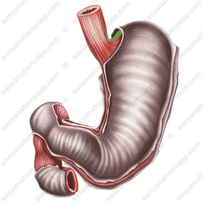
The opening between the esophagus and the stomach is called the cardial orifice (ostium cardiacum).

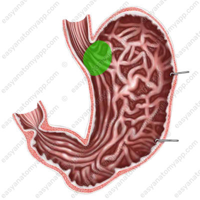
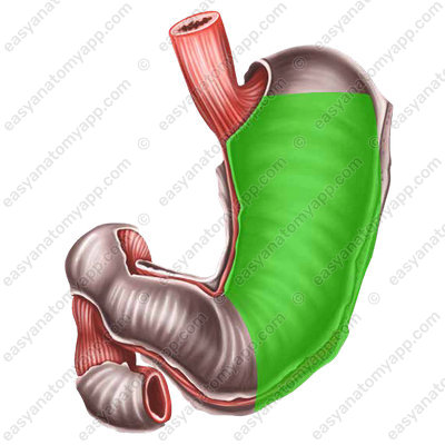
The body of the stomach or the gastric body (corpus ventriculi)
The pyloric part (pars pylorica), which is the narrowed right part of the stomach.
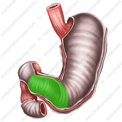
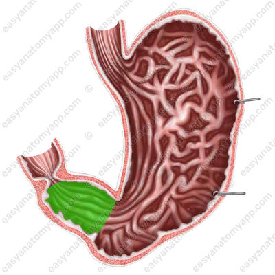
It has a wide part called the pyloric antrum (antrum pyloricum);

and a narrower one, called the pyloric canal (canalis pyloricus), connected to the duodenum.
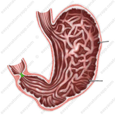
There is a pyloric orifice (ostium pyloricum) between the stomach and duodenum.
The left convex edge of the stomach forms the greater curvature (curvatura major).
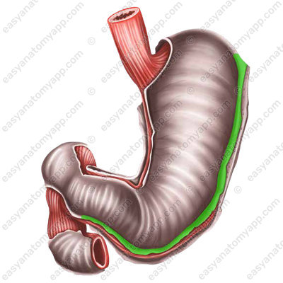
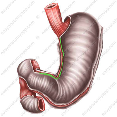
The right concave edge forms the lesser curvature (curvatura minor).
There is a cardial notch (incisura cardialis) between the esophagus and the fundus of the stomach,
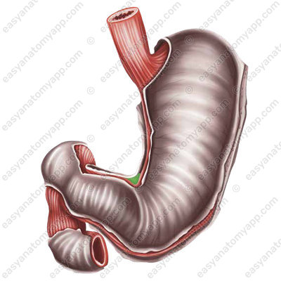
and an angular incisure (incisura angularis) between the body and the pyloric part.
The stomach has two walls:
- The anterior wall (paries anterior)

- The posterior wall (paries posterior)
Both walls pass into one another along the greater and lesser curvatures.
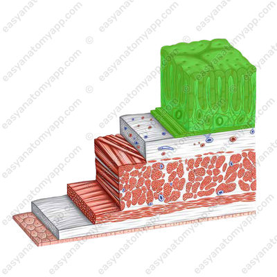
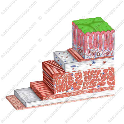
The wall of the stomach consists of several layers:
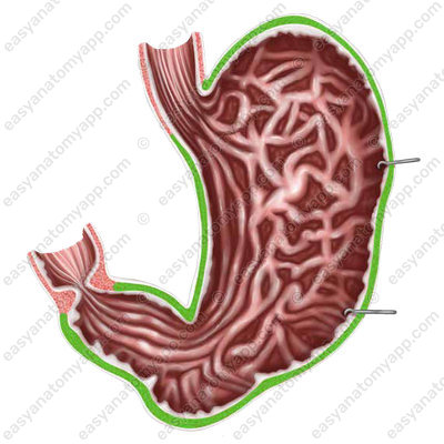
1. Mucous membrane (tunica mucosa)
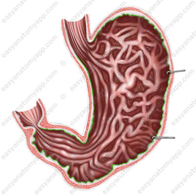
On its surface, it has 4-5 longitudinal folds directed along a lesser curvature from the pyloric orifice. In the area of the pyloric orifice, the mucous membrane forms a circular fold, or the cusp of the pylorus, which, when the pylorus sphincter contracts, completely separates the stomach cavity from the duodenum.
Also, gastric areas (area gastricae) are visible on the surface of the mucous membrane,
as well as depressions between them called gastric pits (foveolae gastricae).
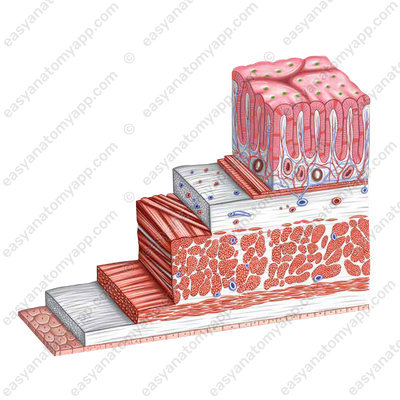
The glands lying in the lamina propria of the mucous membrane open in each pit, producing gastric juice. The mucous membrane of the stomach is covered with a single-layer cylindrical epithelium.
2. The second layer is the submucosa (tela submucosa)

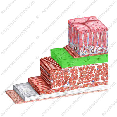
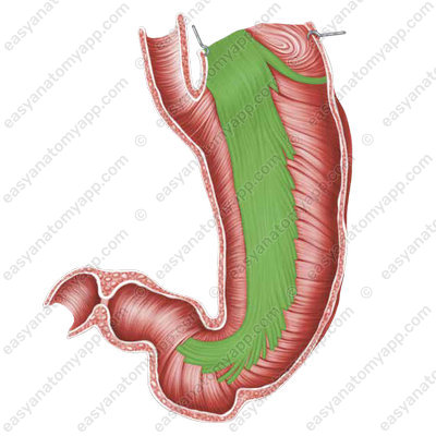
3. Muscular layer (tunica muscularis)
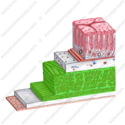
It forms three sub-layers:
- External longitudinal layer (stratum longitudinale)


- Middle circular layer (stratum circulare)

- Internal oblique layer (stratum obliquae). In the area of the transition to the duodenum, this layer thickens, forming the pyloric sphincter
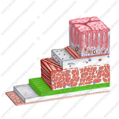
4. The outermost layer is the serosa or peritoneum (tunica serosa), which covers the stomach intraperitoneally.

It is important to understand how the stomach is located in relation to neighboring structures.
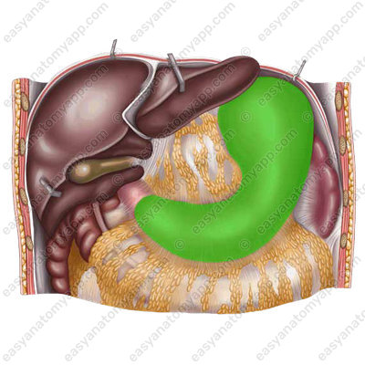
- It is important to understand how the stomach is located in relation to neighboring structures.
- The cardial part, the fundus and the body of the stomach are in contact with the diaphragm
- The lesser curvature is in contact with the visceral surface of the left lobe of the liver
- A small triangular area of the body of the stomach adhere to the anterior abdominal wall.
- The omental bursa, located behind the stomach, separates it from the organs located retroperitoneally.
- The posterior surface of the stomach in the area of the greater curvature adheres to the transverse colon and its mesentery, and the fundus of the stomach adheres to the spleen.
- Posteriorly to the body of the stomach, the superior pole of the left kidney and the adrenal gland, as well as the pancreas are located retroperitoneally.
Physiology of stomach
The activity of the muscles of the stomach determines its motility, maintains tone, almost stable pressure in the lumen of the stomach and performs mixing and emptying. Mixing in the stomach occurs due to peristalsis, which begins in the upper part, in the region of the cardia, and goes towards the pylorus; the interval between contractions is about 20 seconds.
The gastric glands produce 2-3 liters of gastric juice. As a result of mixing food masses with gastric juice, chyme is formed, which is a semi-fluid mass that is expelled from the stomach in separate portions after digestion. Carbohydrates are excreted from the stomach the fastest, proteins are excreted somewhat slower, and fats are digested the longest. Vagus nerves increase the tone of the stomach, enhance its peristalsis, and regulate emptying.
Blood supply
- The left gastric artery (a. gastrica sinistra) supplies blood from the common hepatic artery (a. hepatica dextra)
- The right gastric artery (a. gastrica dextra) supplies blood from the common hepatic artery (a. hepatica communis)
- The right gastro-epiploic artery (a. gastroepiploica dextra) supplies blood from the splenic artery (a. lienalis)
- The left gastro-epiploic artery (a. gastroepiploica sinistra) supplies blood from the splenic artery (a. lienalis)
- Short gastric arteries (a. gastricae breves) supply blood from the splenic artery (a. lienalis)
Venous drainage
Through the eponymous veins
Lymph drainage
Regional lymph nodes
- Left gastric lymph nodes (nodi lymphatici gastric sinistri)
- Right gastric lymph nodes (nodi lymphatici gastric dextri)
- Pyloric lymph nodes (nodi lymphatici pylorici)
- Prepyloric lymph nodes (nodi lymphatici prepylorici)
- Right gastro-omental lymph nodes (nodi lymphatici gastroomentales dextri)
- Left gastro-omental lymph nodes (nodi lymphatici gastroomentalis sinistri)
- The cardial lymphoid ring (anulus lymphaticus cardiae)
Distant lymph nodes
- Splenic lymph nodes (nodi lymphatici lienales)
- Hepatic lymph nodes (nodi lymphatici hepatici)
- Left pancreaticoduodenal lymph nodes (nodi lymphatici pancreatoduodenales sinistri)
- Left supraclavicular lymph nodes (nodi lymphatici supraclaviculares)
Innervation
The vagus nerve (n. vagus) and sympathetic nerves of the coeliac plexus (plexus coeliacus) participate in the formation of the gastric plexus
Anatomy of the stomach
- Stomach
- ventriculus
- Cardial part
- pars cardiaca
- Body of the stomach
- corpus ventriculi
- Greater curvature
- curvatura major
- Lesser curvature
- curvatura minor
- Cardial orifice
- ostium cardiacum
- Pyloric part
- pars pylorica
- Pyloric antrum
- antrum pyloricum
- Pyloric canal
- canalis pyloricus
- Anterior wall
- paries anterior
- Posterior wall
- paries posterior
- Mucous membrane
- tunica mucosa
- Gastric areas
- area gastricae
- Gastric pits
- foveolae gastricae
- Submucosa
- tela submucosa
- Muscular layer
- tunica muscularis
- Longitudinal layer
- stratum longitudinale
- Circular layer
- stratum circulare
- Oblique layer
- stratum obliquae
- Serous coat
- tunica serosa

