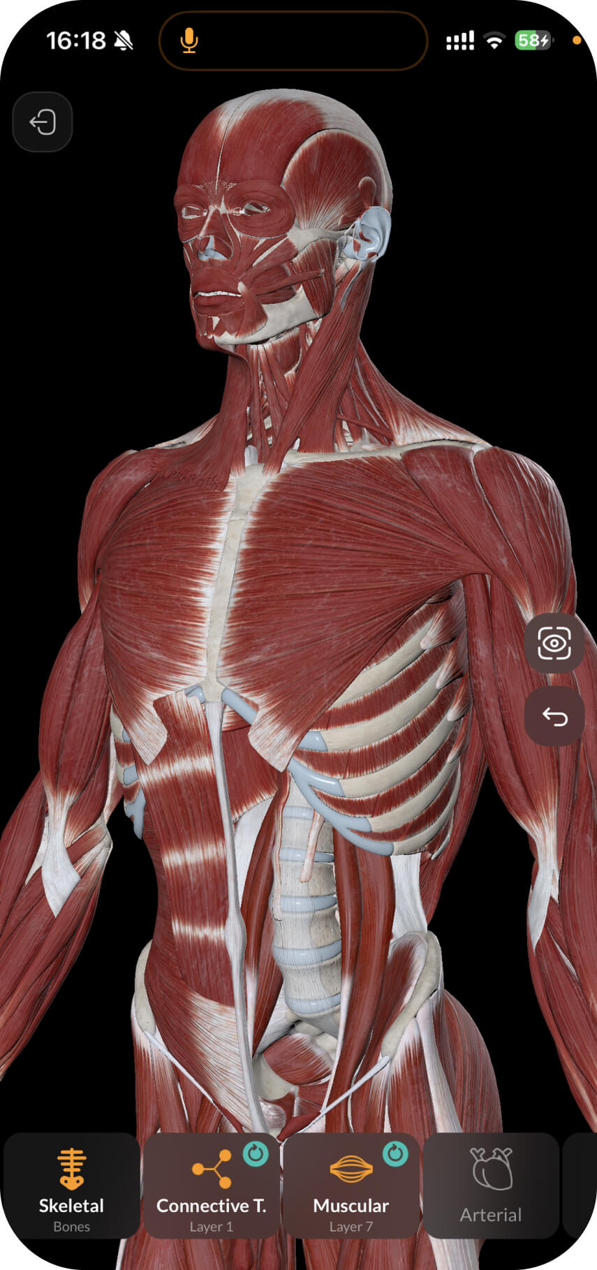
In this note, we will consider the anatomy of the palate (palatum).
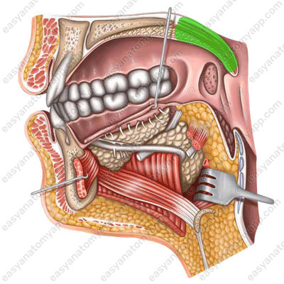
It forms the roof of the oral cavity and is composed of two parts: the hard palate (palatum durum) and the soft palate (palatum molle).
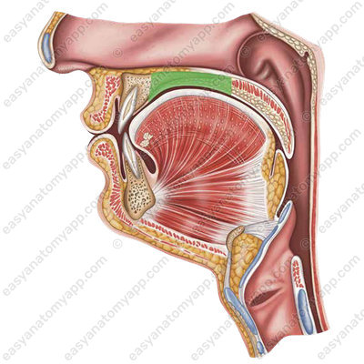

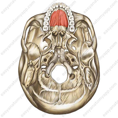
The basis of the hard palate are the palatine processes of the maxillary bones and the horizontal plates of the palatine bones.

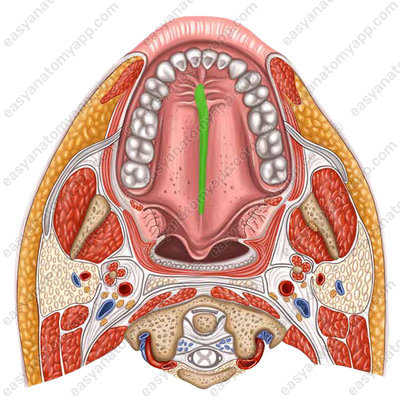
There is the palatine raphe (raphe palati) on the mucous membrane along the midline.
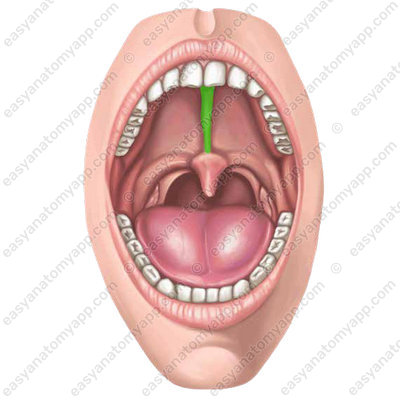
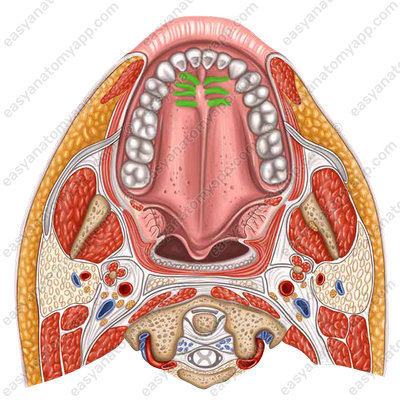
Several transverse folds or palatine rugae (plicae palatinae transversae) diverge from it in both directions.
The soft palate is formed by the palatine aponeurosis and the muscles of the soft palate, which are woven into this aponeurosis.
The unattached posterior border of the palate is called the soft palate or velum (velum palatinum).
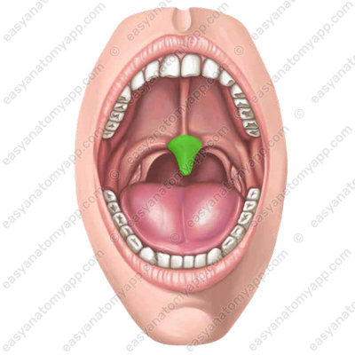
It has a small, rounded process called the uvula (uvula palatina).
Two arches start from the lateral margins of the soft palate.
The palatoglossal arch (arcus palatoglossus), which descends to the lateral surface of the tongue.
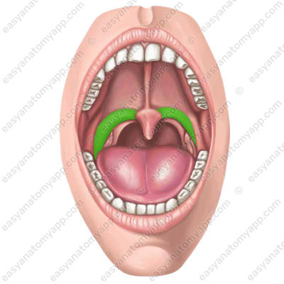
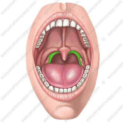
The palatopharyngeal arch (arcus palatopharyngeus), which is directed inferiorly to the lateral wall of the pharynx.
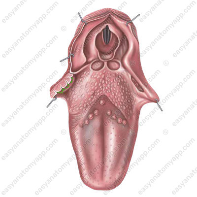
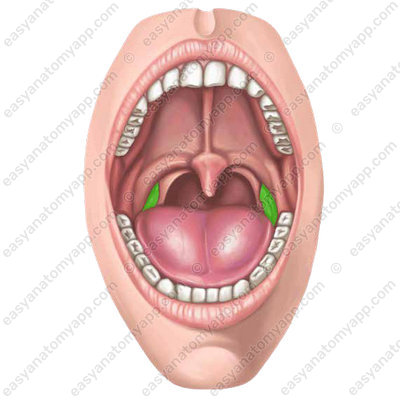
Between both arches on each side, there is a tonsillar fossa or tonsillar sinus (fossa tonsillaris), in which the palatine tonsil (tonsilla palatina) is located.
Let’s look at the muscles of the soft palate.
The tensor veli palatini muscle (m. tensor veli palatini), which arises from the cartilaginous part of the auditory tube and the spine of the sphenoid bone. It weaves into the aponeurosis of the soft palate. It tenses the soft palate and dilates the lumen of the auditory tube.
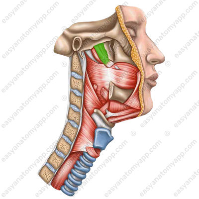
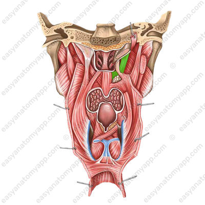
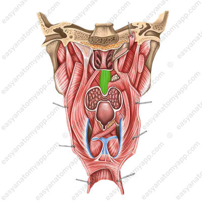
The levator veli palatini muscle (m. levator veli palatini), which arises from the inferior surface of the petrous part of the temporal bone and on the cartilaginous part of the auditory tube. It weaves into the aponeurosis of the soft palate. The contraction of this paired muscle raises the soft palate superiorly, which prevents food from entering the nasal cavity during swallowing.
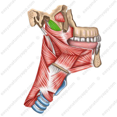
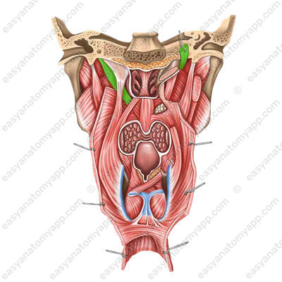
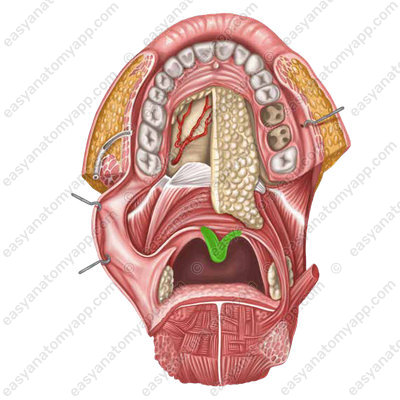
The musculus uvulae (m. uvulae), which is the only unpaired muscle . It arises from the posterior nasal spine and the palatine aponeurosis. It weaves into the mucous membrane of the uvula. The contraction of this muscle raises and shortens the uvula.
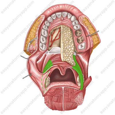
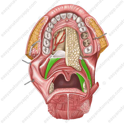
The palatoglossus muscle (m. palatoglossus) and palatopharyngeal muscle (m. palatopharyngeus), which are located deep into the eponymous arches. The contraction of these muscles lowers the soft palate inferiorly, reducing the size of the fauces.
Blood supply
The ascending palatine artery (a. palatina ascendens), which is a branch of the facial artery (a. facialis), supplies the soft palate with arterial blood.
Venous drainage
The venous blood is drained into the pterygoid venous plexus (plexus venosus pterygoideus). The sensory innervation is provided by the lesser palatine nerve (n. palatinus minor) and the pharyngeal branches of the glossopharyngeal nerve (nervus glossopharyngeus).
Innervation
The greater palatine nerve (n. palatinus major), which is a branch of the facial nerve (n. facialis), is responsible for gustatory sensitivity.
Motor innervation is provided by the vagus nerve (n. vagus). Only the tensor veli palatini muscle is innervated by the mandibular nerve (n. mandibularis)
Parasympathetic innervation of the salivary glands is carried out by the otic ganglion (g. oticum).
Hard and soft palate
- Palate
- palatum
- Hard palate
- palatum durum
- Palatine raphe
- raphe palati
- Transverse folds
- plicae palatinae transversae
- Soft palate
- palatum molle
- Tensor veli palatini muscle
- m. tensor veli palatini
- Levator veli palatini muscle
- m. levator veli palatini
- Palatoglossus muscle
- m. palatoglossus
- Palatopharyngeus muscle
- m. palatopharyngeus
- Musculus uvulae
- m. uvulae
- Velum
- velum palatinum
- Palatoglossal arch
- arcus palatoglossus
- Palatopharyngeal arch
- arcus palatopharyngeus
- Tonsillar fossa
- fossa tonsillaris

