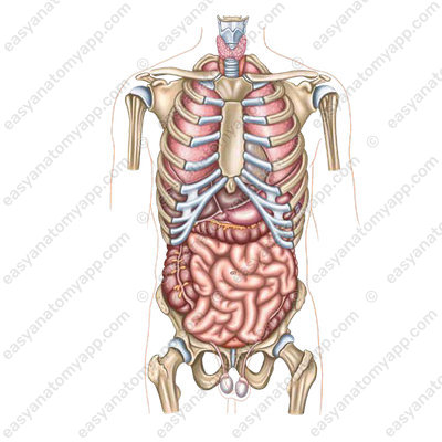
Splanchnology is the study of internal organs.
They are located inside the cavities of the body, differ in shape and size, and perform certain functions.
According to their internal structure, the organs of the digestive system are divided into three groups:
- Tubular organs
- Parenchymal organs
- And organs with a specific structure
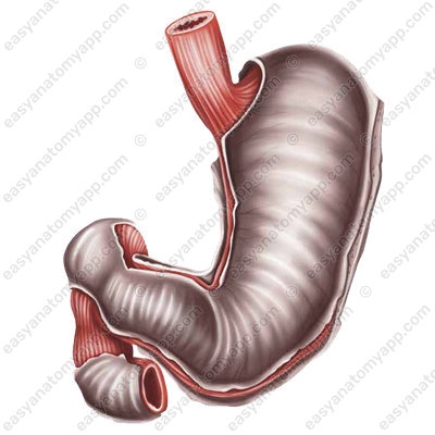
Tubular or hollow organs have a similar wall structure and contain a cavity. Such organs include:
- Esophagus
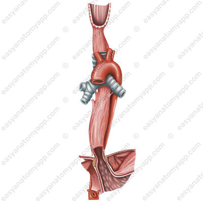
- Stomach
- Small intestine
- Large intestine

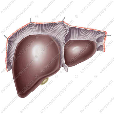
- Gallbladder

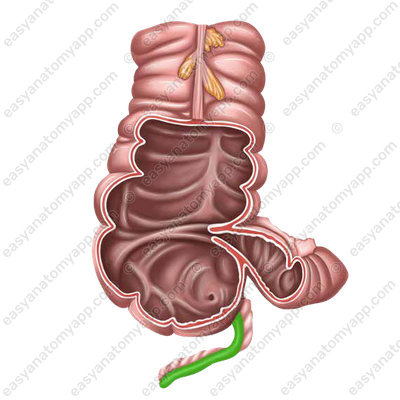
- Appendix
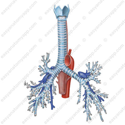
- Trachea and bronchi
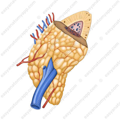
Parenchymal organs include:
- Liver
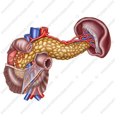
- Pancreas
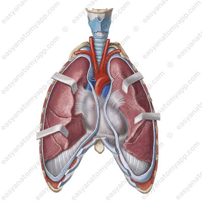
- Lungs
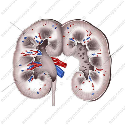
- Kidneys
- Lymph nodes

- Thymus
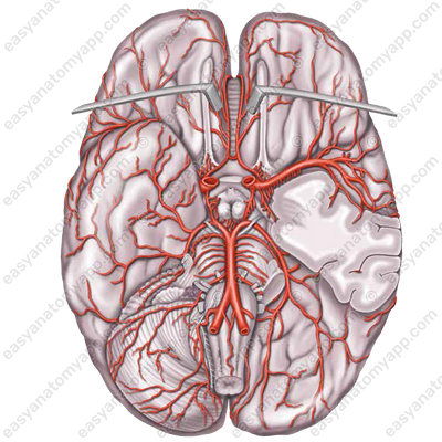
- Brain
- Adrenal glands
- Ovaries

- Testicles

They are composed of a mass of the same consistency.
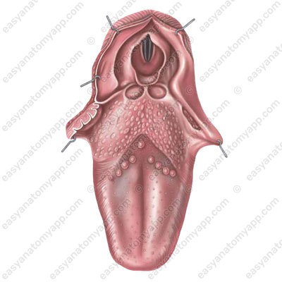
Only a few organs differ in the specific features of their structure. These include the:
Tongue, which is composed of a mucosal membrane and muscles
and the teeth, which are composed of hard tissues.

Each organ is described according to the following parameters:
- Holotopy, which is the location of an organ in a specific cavity or area of the human body. In this case, the organ is projected onto the surface of the body.
- Skeletotopy, which is the location of an organ in relation to certain bone structures.
- Syntopy, which is the location of an organ in relation to other organs.
- Macroscopic structure of an organ, which includes its main components, surfaces, borders, etc.
- Blood supply to the organ, which includes the names of the vessels through which blood flows to the organ and away from it
- Innervation of an organ, which includes nerves innervating an organ.
- Lymph outflow, which includes the main lymph nodes and lymph outflow pathways.
Parenchyma is the main tissue of organs surrounded by connective tissue called stroma.
Vessels and nerves pass through the stroma.
Structural and functional units are the smallest parts of parenchymal organs capable of performing their function, limited by a connective tissue framework with its proper vascular bed.
A segment is a macroscopically visible part of an organ that has relatively autonomous blood circulation, lymph circulation and innervation.
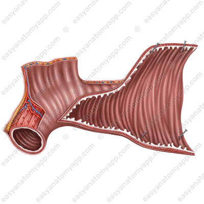
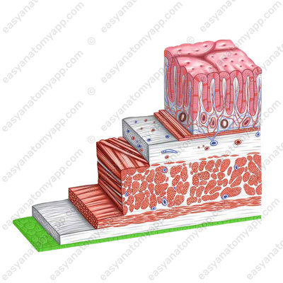
Tubular organs have three tunicae as part of their walls:
- Mucosa (tunica mucosa)
- Muscular layer (tunica muscularis)
- Adventitia (tunica adventitia)
The mucosa (tunica mucosa) lines the internal surface of hollow organs. Its description reflects the features of the epithelium. It can be either a multi-layered epithelium (of cutaneous type, as in the oral cavity) or a single-layered epithelium (as in the stomach or intestines). The nature of the folds (longitudinal, transverse) is also noted.
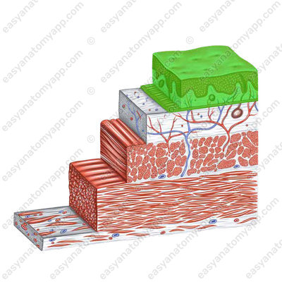
The submucosa (tela submucosas) lies on the border of the mucous and muscular membranes. If the folds of the organ are prominent, that means that the submucosa is well developed. It is structurally defined as a connective tissue in which the vascular and nerve plexuses are located. It has high mechanical strength.
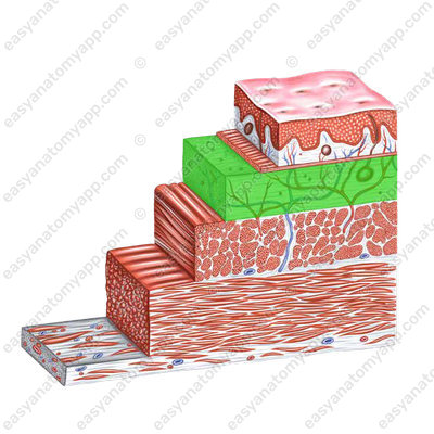
The muscular coat is the middle sheath of a hollow muscular organ. In most cases, it is represented by two differently oriented layers of smooth muscle tissue.
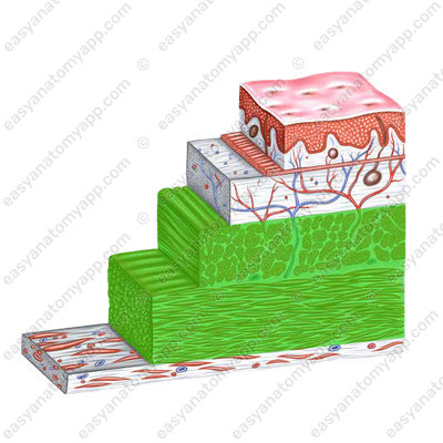
The circular layer (stratum circulare) is located inside, directly behind the submucosa, thickens in a number of organs with the formation of sphincters.
The longitudinal layer (stratum longitudinale) lies externally.
The external sheath of hollow organs may be composed of:
- Adventitia (tunica adventitia)
- Serosa (tunica serosa)
The adventitia (tunica adventitia) is a part of organs that fuse with the surrounding tissues: pharynx, esophagus, duodenum, etc. They cannot move because they are tightly fixed to neighboring structures.
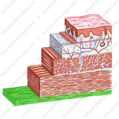
The serosa (tunica serosa) is a thin, transparent plate, its basis is a fibrous connective tissue covered from the outside with a layer of flat cells. It is called mesothelium. It is capable of producing and absorbing serous fluid. Serous fluid reduces friction when the shape and position of the organ changes.
Organs covered with the serous coat have some mobility and can shift.
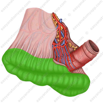
The peritoneum (peritoneum) is the serous coat covering most of the abdominal organs.

Anatomy of the viscera
- Digestive system
- systema digestorium
- Lip
- labium
- Oral cavity
- cavitas oris
- Cheeks
- buccae
- Tongue
- lingua
- Pharynx
- pharynx
- Esophagus
- oesophagus
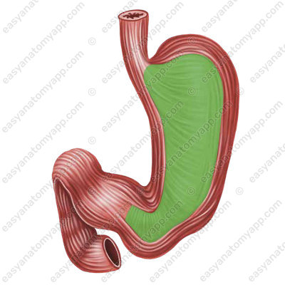
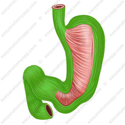
- Stomach
- ventriculus/gaster
- Duodenum
- duodenum
- Liver
- hepar
- Pancreas
- pancreas
- Gallbladder
- vesica fellea
- Jejunum
- jejunum
- Ileum
- ilium
- Caecum
- caecum
- Vermiform appendix
- appendix vermiformis
- Ascending colon
- colon ascendens
- Transverse colon
- colon transversum
- Descending colon
- colon descendens
- Sigmoid colon
- colon sigmoideum
- Rectum
- rectum
- Internal organs
- viscera/splanchna
- Mucous membrane
- tunica mucosa
- Muscular layer
- tunica muscularis
- Adventitia
- tunica adventitia
- Submucosa
- tela submucosa
- Circular layer
- stratum circulare
- Longitudinal layer
- stratum longitudinale
- Peritoneum
- peritoneum

