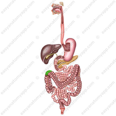
In this note, we will consider the anatomy of the large intestine or large bowel (intestinum crassum).
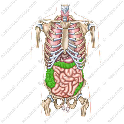
It is located in the abdominal cavity and in the lesser pelvis. The function of the large intestine is the final absorption of fluid from the food bolus, as well as the formation and excretion of feces.
The large intestine consists of several parts.
- Caecum (caecum)
- Ascending colon (colon ascendens)

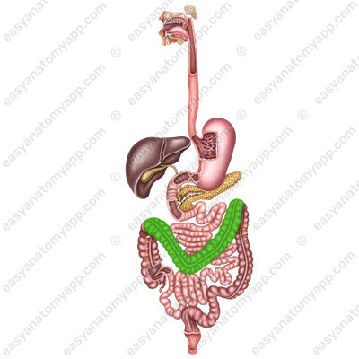
- Transverse colon (colon transversum)
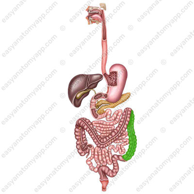
- Descending colon (colon descendens)
- Sigmoid colon (colon sigmoideum)

- Rectum (rectum)

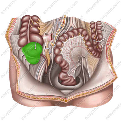
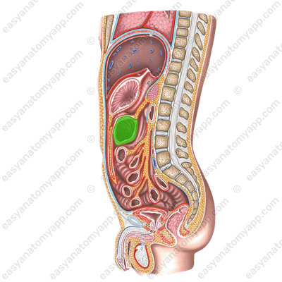
The caecum (caecum) is located in the right iliac fossa, completely covered with peritoneum, and has no mesentery.
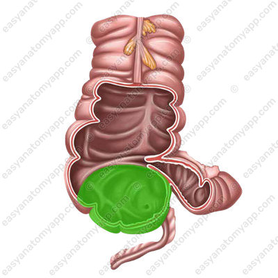
The ileocecal orifice (ostium ileocecale) is located between the ileum and the cecum.
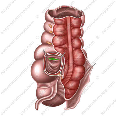
It is covered with folds forming the ileocecal valve (valva ileocaecalis), which is also called Bauhin’s valve.

This valve allows the food bolus to pass toward of the cecum, and not vice versa.
On the posteromedial surface of the cecum, a vermiform appendix, which is a blind lymphoid pouch, arises from the lower wall.

It is completely covered with peritoneum (located intraperitoneally).
In the diagram, you can see various options for the position of the appendix.
The projection of the appendix on the anterior abdominal wall is located on the border of the inferior and middle thirds of an imaginary line drawn between the right anterior superior iliac spine and the umbilicus. This is the so-called McBurney’s point
Ascending colon (colon ascendens) passes to the transverse colon, and forms the right colic (or hepatic) flexure (flexura coli dextra).
From behind, it adheres to the quadratus lumborum muscle and transverse abdominal muscle, to the anterior surface of the right kidney and to the psoas major muscle, from the front, it adheres to the anterior abdominal wall, medially, it comes in contact with the loops of the ileum, and laterally, it comes in contact with the right wall of the abdominal cavity. It is covered with peritoneum from the front and sides (located mesoperitoneally).
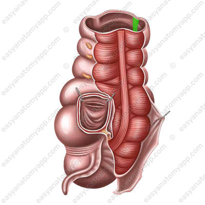
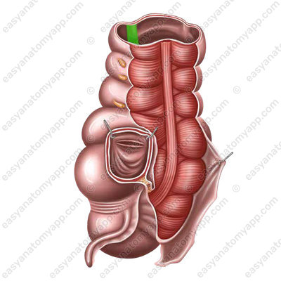
And the transverse colon (colon transversum), passing into the descending colon, forms the left (splenic) flexure of the colon (flexura coli sinistra).

The position of the transverse colon is very variable and depends on the length of the intestine, body type, age. The transverse colon is more often located in the form of an arch, the bulge of which is directed inferiorly. The transverse colon is covered with peritoneum on all sides (located intraperitoneally) and has a mesentery.
From above, the liver and stomach are attached to the right flexure, the spleen is attached to the left flexure, the loops of the small intestine are attached to the left flexure from below, and the duodenum and pancreas are attached to the left flexure posteriorly. When the stomach is empty, it adheres to the anterior abdominal wall, and the filled stomach pushes it down, and it moves away from the abdominal wall.
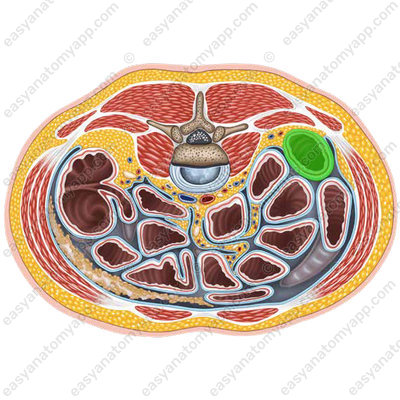
Descending colon (colon descendens) begins from the left colic flexure, goes down and reaches the level of the left iliac fossa, where it passes into the sigmoid colon. It is located in the left part of the abdominal cavity.
The posterior surface of the intestine is attached to the quadratus lumborum muscle, the inferior pole of the left kidney and to the iliac muscle in the left iliac fossa. The anterior surface is in contact with the anterior abdominal wall, the loops of the small intestine are located on the right, and the left abdominal wall is located on the left. The peritoneum covers the descending colon anteriorly and laterally (mesoperitoneal position).
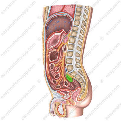
Sigmoid colon (colon sigmoideum) originates from the descending colon at the level of the left iliac fossa.

It passes from the level of the iliac crest to the sacroiliac joint, at the level of which it continues into the rectum.
The intestine has the form of two loops, the shape and size of which are subject to significant individual variations. It is covered with peritoneum from all sides (located intraperitoneally), has a mesentery that attaches to the posterior abdominal wall. The mesentery provides significant mobility.
The anatomy of the rectum is discussed in detail in a separate note.
The large intestine has a number of distinctive features by which we can differentiate it from the small intestine.
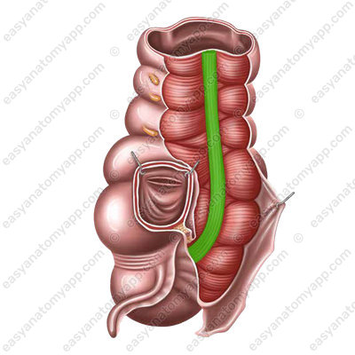
Taeniae (taeniae) are local thickenings of the longitudinal muscular layer
- Omental taenia (taenia omentalis), which is the site of attachment of the omentum
- Taenia libera or free taenia (taenia libera), which is located on the anterior side of the ascending and descending colon and on the inferior side of the transverse colon
- Mesocolic taenia (taenia mesocolica) is the site of attachment of the mesentery
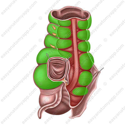
- Haustra (haustrae) are alternating dilations of the lumen of the intestine. They are formed as a result of the discrepancy between the length of the taenia and the parts of the colon between them
- Epiploic appendages (appendices epiploicae) are local deposits of adipose tissue
- The small intestine has a pink color, and the large intestine has a gray color
The wall of all parts of the colon consists of several layers.
- Mucous membrane (tunica mucosa) – consists of a single-layered prismatic epithelium.
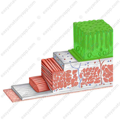
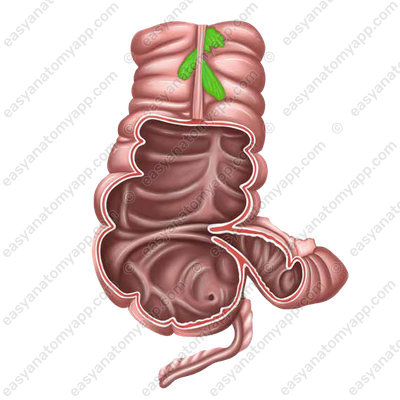
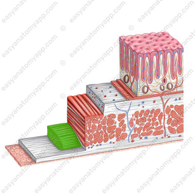
- Submucosa (tela submucosa) is well expressed and forms semilunar folds (plicae semilunares)
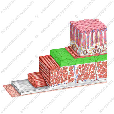
- Muscular layer (tunica muscularis) has two sub-layers.
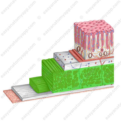
Internal circular layer (stratum circulare)

External longitudinal layer (stratum longitudinale)
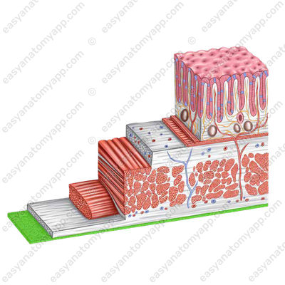
The last, external layer, is the serosa (tunica serosa) or adventitia (adventitia).
Blood supply
- Ileocolic artery (a. iliocolica)
- Right colic artery (a. colica dextra)
- Arc of Riolan (arcus Riolani)
- Left colic artery (a. colica sinistra)
- Inferior mesenteric artery (a. mesenterica inferior)
Venous drainage
Through the eponymous veins to:
- Portal vein (v. portae)
Lymph drainage
- Caecal lymph nodes (nodi lymphatici caecales)
- Paracolic lymph nodes (nodi lymphatici paracolici)
- Sigmoid lymph nodes (nodi lymphatici sigmoidei)
- Mesocolic lymph nodes (nodi lymphatici mesocolici)
Innervation
- Enteric plexus (plexus colicus)
- Vagus nerve (n. vagus)
Anatomy of the large intestine
- Large intestine
- intestinum crassum
- Caecum
- caecum
- Colon
- colon
- Rectum
- rectum
- Taeniae
- taeniae
- Omental taenia
- taenia omentalis
- Free taenia
- taenia libera
- Mesocolic taenia
- taenia mesocolica
- Haustrae
- haustrae
- Omental appendices
- appendices epiploicae
- Mucous membrane
- tunica mucosa
- Submucosa
- tela submucosa
- Plicae semilunares
- plicae semilunares coli
- Muscular layer
- tunica muscularis
- Circular layer
- stratum circulare
- Longitudinal layer
- stratum longitudinale
- Serosa or adventita
- tunica serosa / adventitia
- Ileocecal orifice
- ostium ileocaecale
- Ileocecal valve
- valva ileocaecalis
- Vermiform appendix
- appendix vermiformis
- Ascending colon
- colon ascendens
- Right colic flexure
- flexura coli dextra
- Transverse colon
- colon transversum
- Left colic flexure
- flexura coli sinistra
- Descending colon
- colon descendens
- Sigmoid colon
- colon sigmoideum

